
The rapidly evolving nanotechnology field in cancer researches requires a better understanding of nanoparticles uptake into solid tumors. However, there is not enough information about relationship between nanoparticle properties and nanoparticle transport to the tumor [1]. Nowadays two-dimensional (2D) cell cultures are most frequently used cellular model for the study of cancer biology and treatment. However, a lot of cellular functions and responses that are present in natural tissues are lost in 2D cell cultures. Three-dimensional (3D) cell cultures show a higher degree of morphological and functional similarity to the tissues. Spheroid formation is one of the most well characterized models for 3D cell culture. In cancer research, spheroids could be used as tumor models. Spheroids are self-assembled spherical clusters of cell colonies cultured in environments where cell-cell interactions dominate over cell-substrate interactions, and they naturally mimic avascular tumors with inherent metabolic (oxygen) and proliferative (nutrient) gradients [2].
The aim of our study was to investigate accumulation of quantum dots in 3D spheroid cell culture.
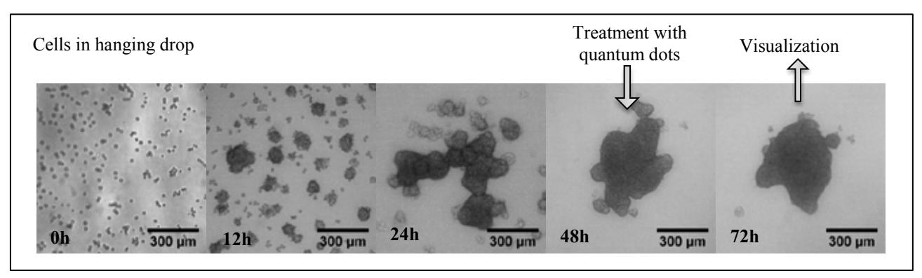
Mouse embryonic fibroblasts (NIH3T3) were used for experiments. 3D cells spheroids were formed using hanging drop technique. NIH3T3 cells were seeded in 96-well hanging drop plate (Biomatrix, USA) and cultivated in 37°C incubator. After 48 h of incubation, 10 % of cell culturing media in each well were changed to a fresh media containing carboxyl quantum dots (Qdot® 625 ITKTM, Invitrogen, USA). The final concentration of quantum dots in each well was 8 nM. After 24 h of incubation with quantum dots, 3D cells spheroids were transferred from hanging drop plate to 8-well microscopy plate. The accumulation of quantum dots were observed using Nikon Eclipse Te2000-U confocal microscope (Nikon, Japan).
3D cell culture at different moments of spheroid formation is represented in Fig. 1. After 24 h of incubation, quantum dots accumulated only in cells which were in the periphery of spheroid. There was not accumulation of quantum dots in the center part of 3D spheroid cell culture. It means that cells do not transfer nanoparticles from one to another. The distribution of quantum dots in each peripheral cell was the same as accumulation of quantum dots in 2D cell culture: nanoparticles accumulated in perinuclear region via endocytosis.
3D spheroid cell culture could be used as avascular tumor model to investigate the uptake and distribution of nanoparticles in the tumor cells. Our results showed that cells in 3D spheroid cell culture do not transfer nanoparticles to each other.