
Metal nanoparticles and nanostructured metal films excited by electromagnetic radiation exhibit collective oscillations of their conduction electrons, known as localized surface plasmon resonance (LSPR), that give these materials a number of interesting and useful optical properties. Possible manipulations of these properties lead to various applications of silver nanoparticles (AgNPs) in optoelectronics [1], biosensing [2], catalysis, metal enhanced optical spectroscopies (metal enhanced fluorescence (MEF) [3], surface enhanced Raman scattering (SERS) [4]), as antimicrobials [5] and other. One of active research fields using AgNPs, which exhibit LSPR in a visible region, is altering emission properties of dyes. For optimized applications of these metal nanostructures and their unique features, it is important to understand the dependence of spectral properties of metal-fluorophore complex on their individual optical behavior.
In this work, layers of AgNPs of different sizes were formed and their influence on absorption and fluorescence properties of low fluorescent conjugated polymer RZ11 and highly fluorescent organic dye Uranin was investigated. Spherical AgNPs were formed by thermal evaporation and annealing: Ag layer was deposited on glass and heated until AgNPs have formed. Samples with AgNPs of approx. 50 nm, 100 nm and 150 nm diameter (which were made from 5 nm, 10 nm and 15 nm of deposited Ag layer respectively) were used for the investigations. Organic materials on layers of AgNPs were spin-coated of few hundred nanometers thickness. Samples were studied by using steady-state and time-resolved absorption and fluorescence spectroscopy.
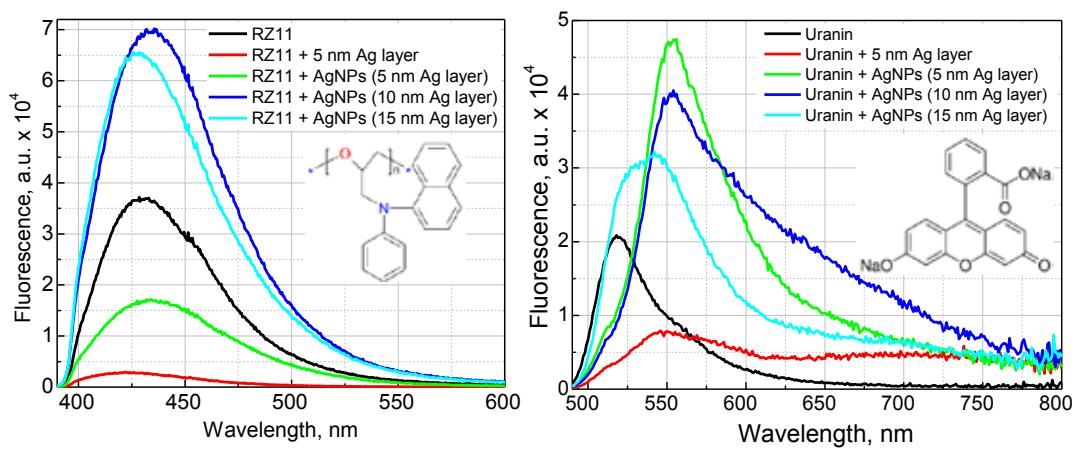
Steady-state absorption spectra showed strongest interaction between fluorophores and AgNPs occurring, when absorption spectrum of organic material overlaps with plasmon resonance band or when it is redshifted compared to the plasmon resonance. Fluorescence studies revealed double fluorescence intensities of organic materials with nanoparticles of approx. 50-150 nm diameter (Fig. 1) and faster decay times than for the samples without nanoparticles. These results indicate fluorescence enhancement effect due to enhanced and confined local electromagnetic fields near the surface of metal nanoparticles and increased radiative decay times. Strongest interaction is observed, when excitation light matches absorption of fluorophore as well as plasmon resonance band. We conclude, that for achieving fluorescence changes or enhancement, absorption and fluorescence spectra of fluorophores and plasmon resonance overlap must be ensured and wavelength of the excitation light is of great importance too.