Magnetic resonance imaging (MRI) is a widely diagnostic imaging method. MRI provides an excellent spatial resolution, but suffers from poor sensitivity. To improve the latter, MRI contrast agents are used. The most commonly used contrast agents are gadolinium-based. Unfortunately, current contrast agents have several disadvantages such as high toxicity or not sufficient enough contrast [1]. That leads to investigation of the novel MRI contrast agents. Gadolinium (Gd3+) has unique paramagnetic properties, therefore it is preferred in development of a next generation MRI contrast agent. In recent years, rare-earth upconverting nanoparticles (UCNPs), which are composed of NaGdF4:Yb,Er, have received significant attention, as they can be useful for several imaging modalities namely MRI (due to the presence of the Gd3+ ion in the host matrix) and optical imaging (due to Yb3+, Er3+ co-doped NaGdF4) [2]. The aim of this study is to evaluate the possibility to use in clinic NaGdF4: Yb,Er nanoparticles as an efficient MRI contrast agents.
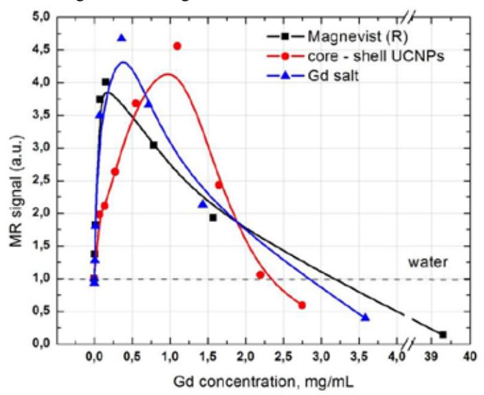
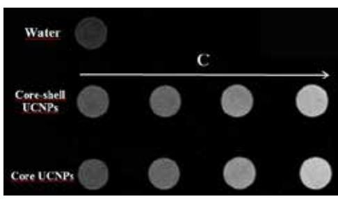
Magnetical properties of TWEEN 80 modified core NaGdF4: Yb,Er and core-shell NaGdF4:Yb,Er@NaGdF4 upconverting nanoparticles were obtained on a 1.5 T Philips Achieva MRI scanner using Sense Flex-M coil. As shown in Fig.1, core and core-shell UCNPs gives a significant positive contrast in the T1-weighted MR images. Significant differences in MR signal enhancement between different types of UCNPs were not observed. Also we investigated MR signal intensity (SI) versus Gd3+ concentration in core-shell UCNPs solution and compared to the clinical contrast agent Magnevist® and Gd3+ salt. Results are shown in Fig.2. In all of the cases, the enhancement in MR signal with the increase of the concentration was obtained until concentration reaches a critical value and the signal begins to decrease. Therefore, it is very important to choose the optimal injection dose in order to obtain the maximum contrast and to avoid signal decreasing effect.
These observations indicate that NaGdF4:Yb,Er upconverting nanoparticles could be applied as an efficient MRI contrast agent. Further investigation is required.