
Nowadays the best-known and the most common biosensing systems are glucose biosensors. They can accurately detect blood free glucose in a short period of time – it is very important for people with diabetes [1]. The active part of the biosensor is glucose oxidase (GOx) enzyme immobilized on surface of the electrode. GOx is a flavin adenine dinucleotide (FAD) containing glycoprotein. FAD is involved in enzymatic redox reactions and determines the enzyme's activity [2]. Glucose oxidase enzyme can exist in several different spatial conformations within its active site FAD can have 'twisted' (stacked) or 'planar' (open) structure [3]. In a planar configuration, flavins have a long lifetime (in nanoseconds), except for the stacked conformation of FAD, where intramolecular electron transfer between the flavin isoalloxazine ring and the adenine moiety takes shorter lifetime periods.
Spectroscopic properties of glucose oxidase (GOx) and flavin adenine dinucleotide (FAD) were investigated. The purpose of this research was to evaluate absorption and fluorescence spectra changes. The steady – state absorption and fluorescence spectra of GOx and FAD are shown in figure 1.
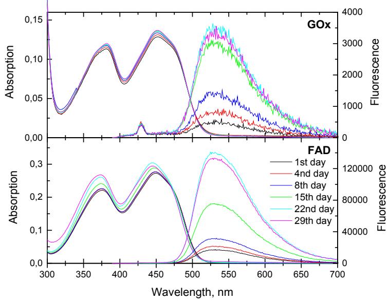
GOx exhibits two absorption bands, which are attributed to FAD absorption bands, with peaks at 380 nm and 450 nm, which are assigned to the $S_2 \leftarrow S_0$ and $S_1 \leftarrow S_0$ and transitions, respectively. These transitions are ascribed to the $\pi-\pi^*$ transition along the three cycles of the isoalloxazine ring in FAD.
GOx fluorescence spectra have two bands at 450 nm and 530 nm. During this study we found that GOx enzyme fluorescence band at 530 nm belongs to coenzyme FAD fluorescence [4]. The increase of GOx fluorescence, which is due to the fact that the enzyme has changed its structure and denatured [2], was visible in 29 day measurement period.
GOx fluorescence lifetimes have been measured at fixed 450 nm and 530 nm wavelengths. The fluorescence decay kinetics were well approximated by a two-exponential model. Average lifetimes of GOx and FAD at 530 nm were coincided and changed from 2,7 ns to 4,5 ns. This confirms that the GOx fluorescence at 530 nm wavelength is determined by FAD fluorescence.