
Nanoparticles (NP) are widely applied in various areas, including food, household chemistry and cosmetics industry. NP have a great perspective in medicine diagnostics and treatment. However, NP are characterized by unique and poorly known nanotoxicity properties [1]. This represents an environmental problem as the quantum dots (QD) may cause toxicity-dependent results, which might be fatal. Regardless of constant increase of NP concentration in environment the danger of contact and possible passage of NP through living organism's protective barriers are not well understood. In general, it is supposed that the chorion protects the embryos from foreign intrusion during development before hatching.
The aim of the present study was to examine of QD passage through a rainbow trout (Oncorhynchus mykiss) embryos chorion and accumulation in embryos structures.
The study was carried out on rainbow trout embryos in "eye-egg" stage. Short-term (2-hour) and long-term (120-hour) exposures and accumulation of QD in embryos structures were performed in climatic camera under static conditions. Embryos were imaged using Nikon ECLIPSE TE2000-U confocal scanning laser microscope equipped with RGB detector with 3 band-pass filters. The 500-530 nm band-pass filter was used for autofluorescence detection and 569-640 nm band-pass filter for QD luminescence detection. Also 4x10⁻⁹ M and 28x10-9 M solutions of CdSe/ZnS-COOH (QD ITK 625) QD with fluorescence maxima at 625 nm were applied during the study. Visualization, analysis, segmentation and interpretation of 2D and 3D structures were managed using "Imaris" imaging software.
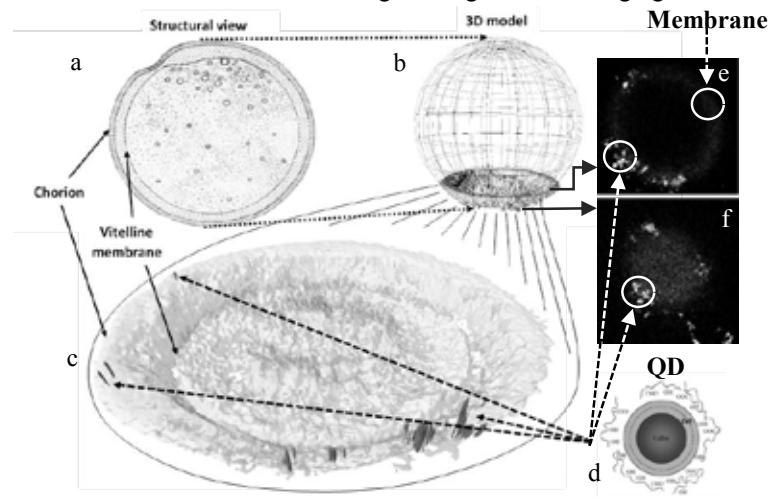
In order to determine accumulation of QD in rainbow trout "eye-egg" stage embryos, incubation of embryos with QD solutions was performed for 2 and 120 hours. To avoid contamination after incubation all embryos were washed using well water. Subsequently, confocal Z-scan imaging of embryos was employed for visualization of embryos at different planes (Fig. 1. e and f). However, we could not see any fluorescence coming from the structures situated deeper than ~ 400 μm. This might occur with regard to weak transparency of embryos. For this reason only a small portion of embryos (~350 μm) was imaged (Fig. 1. b and c). Through the filter 569-640 nm red QD fluorescence was detected (Fig. 1. c, e and f). The green autofluorescence was detected through the filter 500-530 nm. Green color represents autofluorescence of embryos membrane as well as integral and internal proteins (Fig. 1. c, e and f). In 2D images several types of QD distributions, corresponding to different planes of embryos can be observed (Fig. 1. e and f). Nevertheless, 2D images (Fig. 1. e and f) are complicated to clearly understand distribution of QD in membrane. Consequently, we use "Imaris" for better understanding of QD distribution in embryos structures. Likewise 2D images "Imaris" 3D reconstruction showed red fluorescence of QD only in those areas where green autofluorescence was detected (Fig. 1. c, e and f). That is to say, QD adhered to the external surface of chorion of embryos and most likely stuck in it. However, QD did not penetrate through it further to the inner structures (vitelline membrane, yolk and embryo) of the embryos (Fig. 1. c, e and f). In this particular case chorion likely restricted QD uptake. Results approve presumption about chorion protective function against NP.