
This study has been focusing on the development and optimization of a biochip architecture by means of lipid dip-pen nanolithography (DPN). This scanning probe fabrication technique allows direct writing or arraying variety of biomolecules including lipids. As comparedto other molecular inks, lipid DPN displays unusually high process speeds and yields micro/nanopatterns over~mm2 surface areas within minutes. Lipid DPN technique has been successfully tested in screening of cellular membrane receptor-mediated interactions and in studies of protein crystals. Based on these observations we have employed lipid DPN for arraying proteins on pre-structured self-assembled monolayer surfaces.
Protein domain formation was a two-step process. First, structured self-assembled monolayer surface containing hydrophilic (tri-(ethylene glycol)-terminated thiol) and hydrophobic (methyl-terminated eicosane thiol) areas were fabricated using micro-contact printing technique. Surfaces, containing such microdomains were further used to guide lipid spreading during DPN process using 1, 2-dioleoyl-sn-glycero-3-phosphocholine (DOPC) and biotin labeled 1,2-dioleoyl-sn-glycero-3-phosphoethanolamine (Bt-PE) lipid mixture (100:1) as an ink. This process was characterized in a great detail using scanning probe microscopy analysis [1] and revealed the formation of multilamellar lipid assemblies. Lipid patterns remained present during transfer of the sample through the air/water interphase. Finally, lipid structures containing biotinyl functional groups were successfully tested in aqueous environment for selective streptavidin protein immobilization by means of fluorescence microscopy.
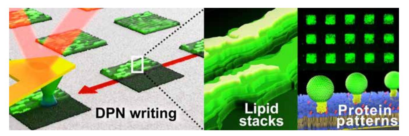
Based on these observations we were able to guide DPN lipid spreading and form lipid domains of pre-defined shape and size for selective protein immobilization. These results promote further development of micro and nano-patterned lipid domains which can be further utilized for lipid membrane mimetics in microarray and lab-on-a-chip device formats.