DIRECTED ASSEMBLY OF MICRO PARTICLES INTO WELL-ORGANIZED ARRAYS
Mindaugas Juodėnas1, Dainius Virganavičius1, Tomas Tamulevičius1, 2, Viktoras Grigaliūnas1, Morten Madsen3, Sigitas Tamulevičius1, 2
1 Institute of Materials Science, Kaunas University of Technology, Lithuania
2 Department of Physics, Kaunas University of Technology, Lithuania
3 Mads Clausen Institute, University of Southern Denmark, Denmark
Recent advances in nanoscience developed elaborated synthesis of nanoparticles of various shapes, sizes and materials. Most applications of nano and micro particles demand their precise stacking into one, two or three dimensional assemblies. Such applications involve novel biosensors [1], nanoelectronic [2], nanophotonic devices [3], drug delivery [4], fluorescence biological labels [5], etc.
One of the techniques for achieving such particle positioning is capillary force assisted assembly. Its basic principle is as follows: a drop of suspension with nanoparticles is confined between a template with surface structures designed for the specific application and a clamping glass slide. These parallel surfaces are translated with respect with each other. Because of evaporation at computer controlled template temperature a particle accumulation zone forms at the three phase contact point i.e. at the edge of liquid meniscus. As it passes over particular size surface structures it deforms and directs particles into predesigned traps. Once the meniscus depins from the structure, the particle stays in the trap. This method offers high arrangement precision, selectivity, throughput and scalability. Several investigations of assembly speed, template temperature and trap geometry have been performed by other authors [6, 7], however the mechanism leading to particle assembly is not quite fully understood.
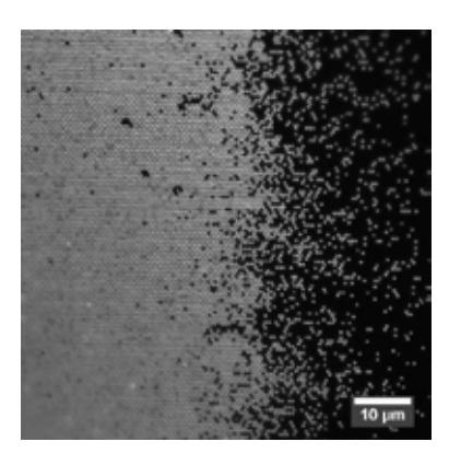
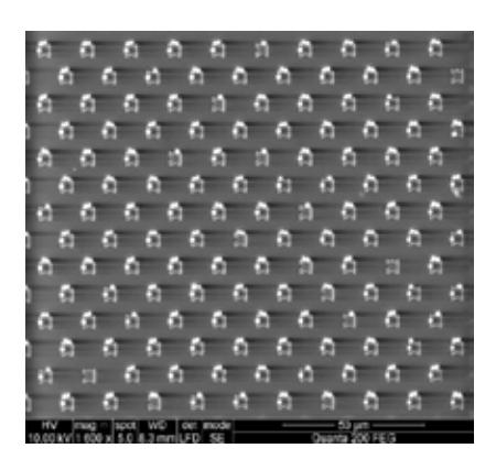
Here we present results achieved employing custom built capillary force assisted particle assembly setup. Holographic and electron beam lithographies in combination with dry etching of crystalline silicon and soft lithography replication in temperature curable polymer were employed to form 4 cm2 area templates. Arrays of fluorescent particles (Fig. 1) and ordinary polystyrene beads (Fig. 2) were assembled into ordered 2D patterns and investigated using optical and scanning electron microscopy. Such regular micro and nano particles arrays are expected to be used for anti-counterfeiting applications and surface enhanced Raman scattering (SERS).
Fig. 1. Fluorescent microscope micrograph of transition zone between assembly areas (left) and non-assembly (right) of 270 nm particles assembled into polydimethylsiloxane (PDMS) template pits of 300 nm diameter. Scale bar 10 μm
Fig. 2. SEM micrograph of 1.5 μm beads assembled into geometric traps on a PDMS template. Scale bar 50 μm
Acknowledgments: Lithuanian-Japan bilateral cooperation partnership programme project (No. LJB-1/2015) was financed by Research Council of Lithuania.
[1] R. L. Edelstein, C. R. Tamanaha, P. E. Sheehan et al., The BARC biosensor applied to the detection of biological warfare agents, Biosensors Bioelectron.2000; 14:805-813.
[2] M. Homberger, U. Simon, On the application potential of gold nanoparticles in nanoelectronics and biomedicine, Phil. Trans. R. Soc. A.2010; 368:1405-1453.
[3] I. Venditti, I. Fratoddi, C. Palazzesi et al., Self-assembled nanoparticles of functional copolymers for photonic applications, Journal of Colloid and Interface Science.2010; 348(2):424-430.
[4] S. Biffi, R. Voltan, E. Rampazzo et al., Applications of nanoparticles in cancer medicine and beyond: optical and multimodal in vivo imaging, tissue targeting and drug delivery, Expert Opin Drug Deliv.2015; 12(12):1847-1849.
[5] M. Bruchez, M. Moronne, P. Gin et al., Semiconductor nanocrystals as fluorescent biological labels, Science.1998; 281:2013-2016.
[6] L. Malaquin, T. Kraus, H. Schmid et al., Controlled particle placement through convective and capillary assembly, Langmuir: The ACS Journal of Surfaces and Colloids.2007; 23(23):11513-11521.
[7] N. Songbo, J. Leemann, H. Wolf et al., Insights into mechanisms of capillary assembly, Faraday Discuss.2015; 181:225-242.