The term vitamin D refers to a group of fat-soluble secosteroids. Two major forms of vitamin D are D2 – ergocalciferol - which is found in plants and yeasts, and D3 – cholecalciferol - which is found in animals and humans. The Vitamin D Receptor (VDR) also known as NR1I1 is a transcription factor. Due to the activation by calcitriol, the VDR and retinoid-X receptors form a heterodimer which changes its conformation. The heterodimer bonds to hormone response elements on DNA, which enables regulation of transcriptional response. Moreover, VDR is involved in microRNA-directed post-transcriptional mechanism which is connected with the influence of calcitriol on human bones. VDR could be find in many tissues, where plays various roles. So vitamin D has a lot of applications including regulation of calcium-phosphate metabolism, antiproliferation action, decreasing possibility of developing tumor and many others.
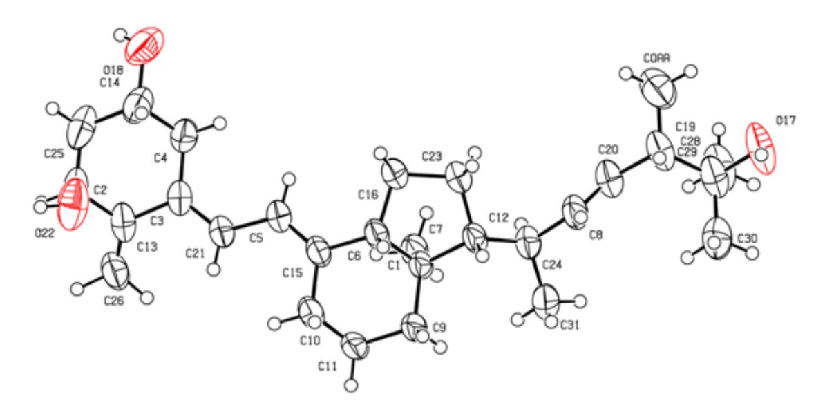
The crystal structures of 1,25-dihydroxyergocalciferol and 1,25-dihydroxycholecalciferol analogues have been determined and the result of this research will be presented. The crystal and molecular structures of these compounds were solved and refined using single crystal X-ray diffraction. Three of analogues crystallize in the P212121 space group of the orthorhombic crystal system with the unit cell parameters: a=9.62082(8) Å, b=14.18309(12) Å, c=31.7987(2) Å; a=6.6492(5) Å, b=9.9013(9) Å, c=38.811(3) Å and a=38.0952(3) Å, b=12.95430(10) Å, c=11.07290(10) Å, respectively. One of analogues (Fig. 1) crystallizes in the C2 space group of the monoclinic system, with the unit cell parameters a= 33.410(2) Å, b=6.3780(4) Å, c=12.2329(8) Å. I will discuss structural differences between the above mentioned analogues and factors influencing these differences.