
ZnO-based materials in the low dimensional regime have attracted considerable attention owing to a wide bandgap of 3.4 eV, large excitonic binding energy of 60 meV and their size-dependent optical and electronic properties, based on the quantum confinement effect of the electronic states. Raman scattering allows investigating the internal properties of material, phonon modes, and electron-phonon interactions. Surface enhanced Raman scattering of light by ZnO nanostructures was previously reported by several groups [1,2]. An increase of Raman scattering for nonresonant excitation was obtained for ZnO NCs coupled with colloidal silver nanoparticles [1], ZnO nanorods and nanocrystals covered with silver clusters by vacuum deposition [2]. The enhanced modes were $A_1$ (LO) and $E_2$ (high). In this work we report on significant surface enhanced nonresonant Raman scattering of light by ZnO NCs coupled with silver nanostructures.
Colloidal ZnO nanocrystals solution in ethanol were synthesized by hydrolysis of zinc acetate (reagent grade) with sodium hydroxide (pure) at 0 °C, as described in more details elsewhere [1]. After the synthesis solution was kept at 55–60 °C for 2 h to promote ZnO particles crystallization. The molar zinc oxide concentration in solution was 0.01 M corresponding to about $10^{-5}$ M in terms of NC concentration. Raman scattering experiments were performed on ZnO NCs deposited by drop-casting onto the glass substrate. As the source of plasmonic enhancement a nanostructured silver thin films were deposited by means of the vacuum evaporation technique. Two sorts of samples were prepared: with silver film deposited on glass before ZnO NCs casting or by deposition of the nanostructured metal film on top of the ZnO NCs preliminary applied onto the pure glass surface. In both cases, deposition of ZnO NC film was performed by dropping a 10 µL of ZnO NCs solution onto the substrate and drying for at least 1 hour. The reference sample was prepared in the same way but without metal deposition. Raman spectra were collected in backscattering geometry using Jobin-Yvon Labram confocal spectrometer with He-Ne laser source ($\lambda_{exc}$=632.8 nm).
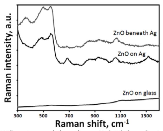
The Raman spectra of the ZnO NCs on and beneath the silver coating are shown in Fig. 1. The most intensive component at about 560 cm-1 coincides with previously obtained mode for ZnO nanocrystals [1,2]. According to the results of Ref.[2], the modes at 490–500 cm-1 and 560 cm-1 can be attributed to the surface optical (SO) phonons formed from phonons of the symmetry $A_1$ or the symmetry $E_1$. Spectrum for ZnO beneath silver nanoparticles shows the additional peak at 373 cm-1, which also have surface nature. The modes in the range 600-1500 cm-1 can be attributed to zinc acetate. Both SERS configurations demonstrated comparable enhancement efficiency and can be used in further experiments.