
Early and accurate detection of cancer might be complicated due to limited efficiency of contrast agents. Gold nanoparticles (AuNP) could be excellent contrast agent for X-ray imaging due to specific properties of gold that overcome some limitations of standard (iodine-based) contrast agents used for imaging in vivo. Gold has higher photoelectric effect cross-section of X-rays, significantly lower clearance rate than iodine and are considered to be nontoxic in living systems [1]. Surgical treatment of cancer might be difficult as well because tumour cells and lymph nodes might be obscure in contrast of other tissues even when contrast agents are injected.
In our study, we synthesized polyvinylpyrrolidone (PVP) stabilized gold nanoparticles, assessed their optical properties and compared pharmacokinetics of synthesized AuNP with standard contrast agent in vivo. Two methods to purify nanoparticles were used and properties of Au-MES-PVP particles were assessed using TEM microscope images.
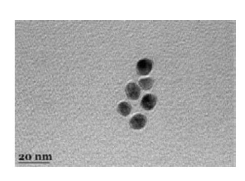
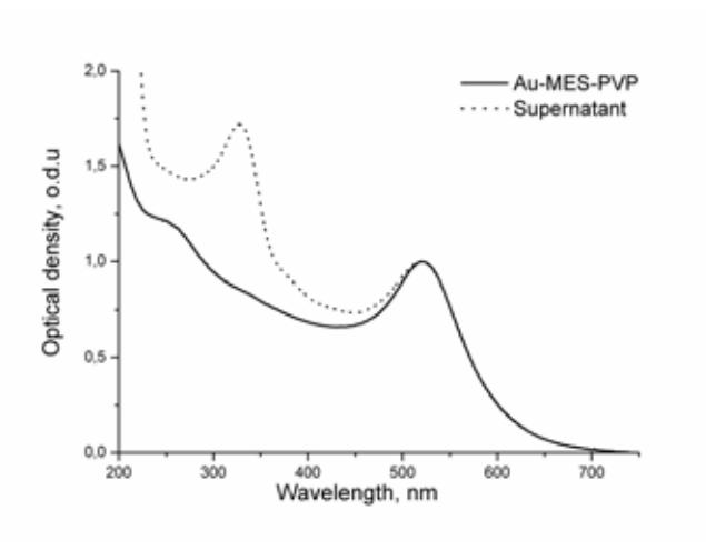
Synthesised AuNP were diluted four times and centrifuged for 1 hour with 10 000 G force, after that the supernatant was removed. The other way that we used to separate particles was with the dialysis diaphragm. Purified with both methods, absorption spectrum of AuNP were similar. According to TEM images (Fig. 1) and absorption spectrum of synthesized AuNP (Fig. 2) [2] they are fairly uniform in size with a diameter of about 15 nm.
To determine pharmacokinetics and X-ray imaging contrast in vivo, BDF1 hybrid male mice were used. One was injected with a clinically approved contrast agent (Ultravist 300) bolus of 0.1 ml directly into the right leg tissue (control sample). The other with the same bolus of synthesized and purified Au-MES-PVP accordingly. X-ray images were acquired before and after the injection at 0, 5, 10, 15, 30, 45, 60 minutes and 24, 48 hours. Initial distribution of contrast agent in the injected tissue were detected in both cases, but after 10 min Ultravist 300 started accumulating in bladder and after 1 hour almost all contrast agent was concentrated there, after 24 hours no traces of Ultravist 300 was noticed. Different pharmacokinetics were observed with Au-MES-PVP nanoparticles. Significant change of X-ray contrast in leg was not observed 1 hour after the injection. But after 24 hours Au-MES-PVP particles concentrated in the lymph nodes on the right side of a mouse and were still seen even after 48 hours. Afterwards, the autopsy of the mouse showed that the lymph nodes on the right side were black and plain seen in the contrast of other tissues.
Results showed that Au-MES-PVP clearance rate is slower than Ultravist 300. Therefore, repeated injections of contrast agents might not be necessary for imaging. Moreover, Au-MES-PVP nanoparticles accumulate in lymphatic system hence it can be used for distinguishing lymph nodes and tissues. Further investigation in this area will provide a deeper level of understanding the properties and possibilities of PVP stabilized nanoparticles in imaging and, perhaps, removing of tumours.