1 Centre for Modern Interdisciplinary Technologies, Nicolaus Copernicus University, Toruń, Poland
2 Institute of Physics, Faculty of Physics, Astronomy and Informatics, Nicolaus Copernicus University, Poland
3 Center for Lifespan Psychology, Max Planck Institute for Human Development, Berlin, Germany
4 University Medical Center Hamburg - Eppendorf, Hamburg, Germany
Several studies have shown that functional network reorganizes from more segregated to more integrated pattern when demands of a cognitive task increase. This pattern of network reorganization was found especially for working memory ($n$-back) task [1-3]. However, differences in network reorganization during a working memory task engaging different modalities and information processing were so far not investigated. Here, we compare how increasing cognitive demands of visuospatial and auditory $n$-back are reflected in the whole-brain functional network topology.
Forty-seven healthy subjects (aged 18-26) were examined using the functional magnetic resonance imaging (fMRI) technique during resting-state and while performing visuospatial and auditory working memory $n$-back tasks at increasing levels of difficulty (from 1-back to 2-back). FMRI data were pre-processed using a standard pipeline and denoised with aCompCor method [4]. We calculated correlation matrices for each participant, task type, and task condition (1-back, 2-back and resting-state) for 264 ROIs functional brain parcellation [5]. For connectivity analysis the Brain Connectivity Toolbox for MATLAB was used [6]. The comparison of strength of individual edges was examined with use of Network-Based Statistic method which is implemented in the NBS Toolbox [7].
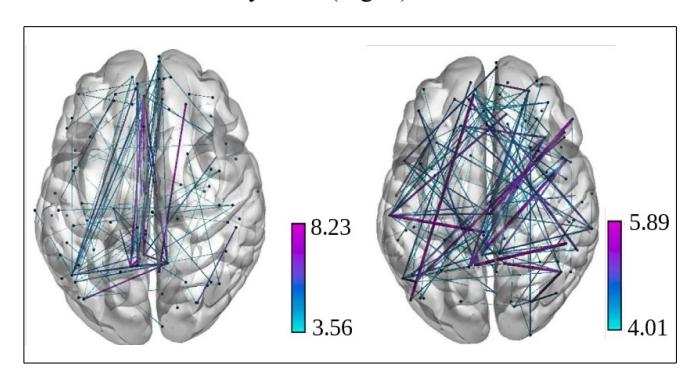
We showed that the brain reorganizes itself while switching from the lower- to the higher-demand task. Both for auditory and for visuospatial tasks, the highest modularity occurred in the resting-state, and the modularity in 2-back was lower than in 1-back condition. However, the modularity during the 1-back auditory task was lower than in the visuospatial 1-back task. NBS analysis showed that for both auditory and visuospatial conditions the majority of weakened network edges consist of edges within the default mode network (DMN) and the largest group of edges in the strengthened network link DMN with other brain systems (Fig. 1).
Fig. 1. Weakened (left, 1-back > 2-back) and strengthened (right, 2-back > 1-back) networks in terms of predefined brain systems while performing visuospatial tasks.
These results confirm the previous findings that the brain adapts to increasing cognitive demands and extend these conclusions for different modalities. Such outcomes can shed new light on the understanding of the differences between various working memory modalities.
Acknowledgments: This project is supported by the National Science Centre, Poland (2015/17/N/HS6/03549; 2017/24/T/HS6/00105).
[1] [1] Braun, U., Schäfer, A., Walter, H., Erk, S., Romanczuk-Seiferth, N., Haddad, L., ... Bassett, D. S (2015), Dynamic reconfiguration of frontal brain networks during executive cognition in humans, Proceedings of the National Academy of Sciences, 112(37), 11678-83.
[2] [2] Finc, K., Bonna, K., Lewandowska, M., Wolak, T., Nikadon, J., Dreszer, J., Duch, W., Kühn, S. Transition of the functional brain network related to increasing cognitive demands. (2017), Human Brain Mapping 38 (7), 3659-74.
[3] [3] Bassett, D. S., Wymbs, N. F., Porter, M. A., Mucha, P. J., Carlson, J. M., & Grafton, S. T. (2011), Dynamic reconfiguration of human brain networks during learning. Proceedings of the National Academy of Sciences, 108(18), 7641-46.
[4] [4] Behzadi, Y., Restom, K., Liau, J., Liu, T. T. (2007), A component based noise correction method (CompCor) for BOLD and perfusion based fMRI. NeuroImage, 37(1), 90-101.
[5] [5] Power J. D., Cohen A. L., Nelson S. M., Wig G. S., Barnes K. A., Church J. A., Vogel A. C., Laumann T. O., Miezin F. M., Schlaggar B. L., Petersen S. E. (2011), Functional network organization of the human brain. Neuron 72, 665-678
[6] [6] Rubinov M, Sporns O (2010), Complex network measures of brain connectivity: Uses and interpretations., NeuroImage 52:1059-69.
[7] [7] Zalesky A, Fornito A, Bullmore ET (2010), Network-based statistic: Identifying differences in brain networks. NeuroImage. 53(4):1197-1207.