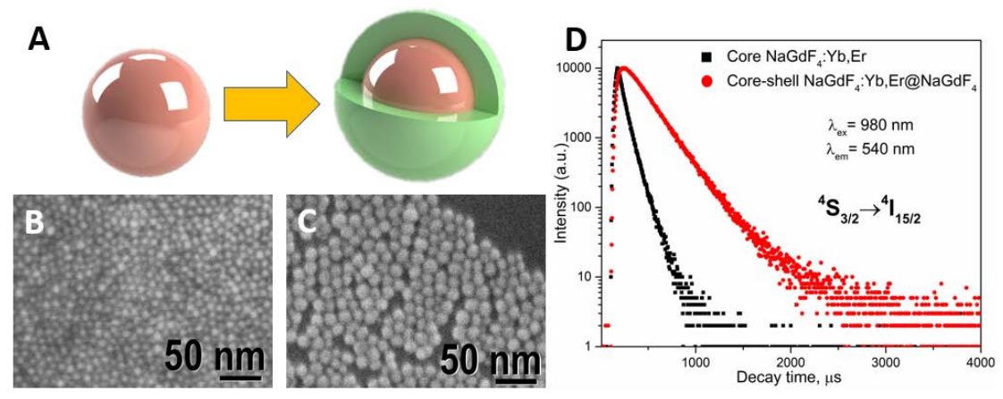
Lanthanide ($Ln^{3+}$) ions doped fluorides nanoparticles have been extensively investigated due to the advantages associated with their unique optical properties. Hexagonal phase $\beta$-NaGdF4 is a promising matrix for the creation optical/magnetic dual-modal bioprobes, but upconversion luminescence (UCL) efficiency of this host material is still low and needs to be improved. A major method to enhance the UCL intensity is to use a core–shell structure, where the inert shell protects the luminescent rare earth ions in the core by passivating the surface lattice defects and shielding the core from the surrounding environment [1]
In this work, we present the thermal decomposition synthesis and surface functionalization of $Ln^{3+}$-doped core-shell upconverting nanoparticles (UCNPs) and demonstrate how they can be used in biological applications and temperature sensing [2]. Yb3+ and Er3+-doped NaGdF4 core-shell nanoparticles with various shell thicknesses of NaGdF4 were successfully prepared. The effect of shell thickness on luminesce intensity and decay kinetics will be presented. X-ray diffraction (XRD), scanning electron microscopy (SEM) and photoluminescence (PL) spectroscopy were used to characterize UCNPs (Fig. 1). The studies show that size and shell thickness have effects on photo-physical properties of core-shell upconverting nanoparticles.