Amyloids are self-assembled, highly ordered, closely packed peptide or protein aggregates and their formation is associated with neurodegenerative diseases, such as Alzheimer's, Parkinson's and prion diseases [1]. Amyloidogenic protein aggregation mechanism dependence on environmental factors, such as ionic strength and pH are still poorly understood [2].
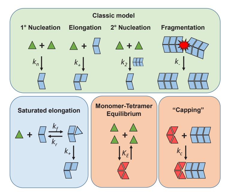
Insulin amyloid-like fibrils were prepared by incubation of fresh insulin solution in 100 mM phosphate buffer pH 2.4 (with and without 0.1 M NaCl) at 60 °C for 24 hours in quiescent conditions. 5.0 mg/ml insulin solutions were used in dynamic light scattering analysis and formed fibrils were examined with atomic force microscopy and infrared spectroscopy. Aggregation kinetics of insulin solutions (0.5 – 5.0 mg/ml) were recorded at constant 60 °C temperature by measuring Thioflavin -T fluorescence intensity. Kinetic data was analysed using "classic" and "saturated elongation" models [3] as well as our proposed "classic" + tetramer and "classic" + "capping" model (Fig. 1.).
In this work we observed unusual insulin aggregation kinetics at pH 2.4 under two ionic strength conditions that could not be explained by any currently predominant models. Our proposed additional tetramer formation and aggregation center "capping" steps greatly improved the "classic" aggregation model's ability to fit the abnormal kinetic data. The addition of salt also shifted the monomer-tetramer equilibrium towards higher oligomeric form creation.