
Among a variety of plasma sources special attention is focused on the development and application of plasma jets [1]due to the possibility to treat objects of complex shapes and sizes outside a closed discharge volume.
The cold plasma jet is formed by direct current glow discharge in air at atmospheric pressure [2]. Interelectrode gap is fixed at 0.7 mm. Discharge is maintained by DC power supply with an output voltage up to 3 kV and ballast up to 80 kOhm. Air flow can be varied in the range of 1-10 l/min. Plasma generated in the discharge is blown out with the gas through anode hole into surrounding air. As a result, a glow of plasma jet of 2-3 mm in diameter and several centimeters in length is observed (Fig. 1, a and b). In almost all articles about plasma medicine the accent is made on the bactericidal action of RONSs (e.g., NO, O3, OH, H2O2, etc.) generated by the plasma jets [3]. Determination of the concentration of long-living chemically active species in the plasma jet is performed by IR absorption spectroscopy [4]. In the absorption spectra of air jet, NO, NO2, HNO2and N2O bands are observed. Molar fractions of these active species at the zone of the jet impact on the bacteria are 40 ppm, 20 ppm, 10 ppm and 2 ppm, correspondingly. The temperature field was measured with a thermocouple and compared with the schlieren images. Three separate temperature regions with well-defined boundaries were observed along the diameter of the jet. The first one is a central narrow hot area that corresponds to the area of the visible luminous jet. It has gas temperature of 30 to 120°C depending on the gas low rate and distance from the anode. It is surrounded by a warm "coat" of diameter of about 1 cm with a temperature of 25 to 50°C (the boundary of this zone is shown in Fig. 1, c). The last region is an ambient air area surrounding the plasma jet and having the room temperature.
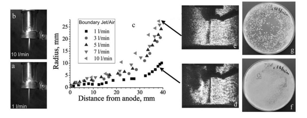
The plasma jet effectiveness was evaluated on test-strains of Staphylococcus aureus with an initial concentration of microorganisms of 106CFU/ml. The temperature in the Petri dish was controlled by a FLIR E4 thermal imager and did not exceed 30 °C. Inactivation ability is demonstrated by inhibition zones (Fig. 1, f and g). At a flow 1 l/min, the inhibition zone is difficult to detect. With increasing gas flow, the zone becomes more pronounced, "clean" regions are formed, and the radius of the zone grows. As it follows from the shadow images, this can be due to the increase in the radius of the plasma jet itself (Fig. 1c).The best effect is observed at 10 l/min, inactivation in this case takes place in the whole Petri dish. Also, as the flow increases, a decrease in the gas temperature of plasma jet in the region of the sample from 50 °C to 35 °C.