
The main purpose of tissue engineering is to develop artificial tissue or organ which could be used for treatment [1]. This study was focused on artificial bone tissue development, because of its necessity in medicine and odontology. The aim of this work was to evaluate the impact of polylactic acid (PLA) scaffolds microstructurization on rat's dental pulp stem cells (DPSC) osteogenic differentiation in vitro.
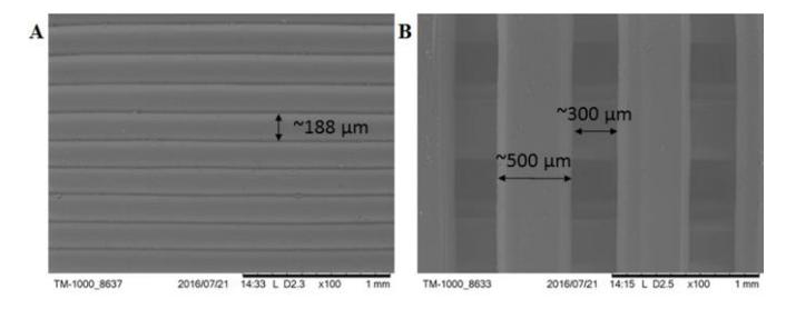
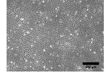
Stem cells were isolated from rat's dental pulp by outgrowth from intact tissue and purified with magnetic beads coated with antibodies against cell surface marker CD44. These cells were immunologically characterized by flow cytometry and their multipotency was examined by osteogenic, myogenic and adipogenic differentiations. PLA scaffolds with two different microstructures were created by 3D printing. Wavy topography consisted of 188 μm wide joined threads, while porous micro-woodpile geometry PLA scaffolds had 300 μm diameter pores. For successful cell number evaluation, new method was established which rely on DAPI fluorescence when it is bound to DNA. This method was used for DPSC proliferation evaluation on PLA scaffolds. Results showed that both microstructurised 3D PLA scaffolds enhanced DPSC proliferation, compared to flat tissue culture polystyrene surface. Osteogenic differentiation results indicated that wavy PLA scaffolds were more suitable for matrix mineralization; however, alkaline phosphatase activity increased the most in the cells, seeded on porous scaffolds. The expression of three osteogenic differentiation genes - Runx2, osteopontin (OPN) and osteocalcin (OCN), were evaluated at different time points using reverse transcription and qPCR. Results showed that Runx2 gene transcription is better activated at beginning stages on wavy scaffold. OPN gene expression is earlier activated on wavy scaffold, however, later on, no difference has been found between two analysed scaffolds. OCN gene expression, which indicates late osteogenic differentiation, wasn't detected. Moreover, it was showed, that cell's environment and scaffolds microstructurization is sufficient enough to induce DPSC osteogenic differentiation.
All things considered, PLA scaffolds are suitable for rat's DPSC proliferation and osteogenic differentiation. Moreover, surface topography influenced DPSC osteogenic differentiation capacity. Ordered scaffolds can be successfully applied as templates for cell proliferation and differentiation.