
Surface enhanced Raman spectroscopy (SERS) is a powerful vibrational spectroscopy technique that allows highly sensitive structural detection of low concentration analytes and is applicable for studies of model systems on roughened electrodes [1]. The active area of our SERS substrates is formed using an ultra-short pulse laser on a soda-lime glass substrate. The resulting surface structure features stochastic nanopattern, which meets good resonance characteristics for various excitation wavelengths and adsorbed analyte molecules. The tethered lipid bilayers (tBLMs) (Fig 1.) are considered as perspective experimental platforms for membrane biosensors and may be suitable for a broad spectrum of biophysical experiments such as peptide/membrane interactions, protein/membrane interactions, lipid phase transitions and others. Attached to a SERS active surface, tBLMs may allow monitoring of biologically relevant events with Raman spectroscopy. It is known that structure of the self-assembled monolayers (SAMs) used to anchor phospholipid bilayers to surfaces affects the functional properties of the tethered bilayer membranes (tBLMs) [2].
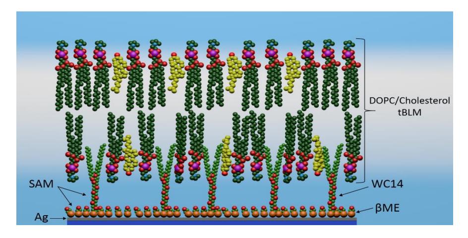
The aims of this study are to test new surface enhanced Raman scattering (SERS) substrates coated with silver to determine their suitability to analyse self-assembled monolayer (SAM) of anchor molecules for tBLM formation on roughed surface. Two types of anchor molecules were chosen to form SAMs on nanostructured silver surface - long strand thiolipid Wilma's compound (WC14) and different short strand back fillers - 2-mercaptoethanol (2-ME), 3-mercapto-1-propanol, 4-mercapto-1-butanol. We received a good quality SERS spectra form SAM of anchor molecules before and after tBLM formation.