Restriction endonucleases (REases) recognize specific nucleotide sequences in a double stranded DNA and cleave both strands of the duplex. In vivo REases protect their host bacteria from viral attacks by cleaving foreign DNA. In vitro, they are widely used as a molecular tool for various DNA manipulations [1]. Restrictase BfiI belongs to the phospholipase D (PLD) superfamily and does not require metal ions for DNA cleavage. It recognizes an asymmetric DNA sequence, 5'-ACTGGG-3', and cuts top and bottom strands at fixed positions downstream of this sequence [2].
Here we are studying several versions of BfiI REase: WT BfiI, BfiI-SS, BfiI-K107A, BfiI-K107A-SS. The wild-type BfiI is a native protein. In BfiI-SS protein SS bridge is linking the dimer interface [3]. In BfiI-K107A protein mutation was introduced in the active center at residue 107 - Lysine was changed to Alanine. BfiI-K107A-SS protein contains both mentioned mutations.
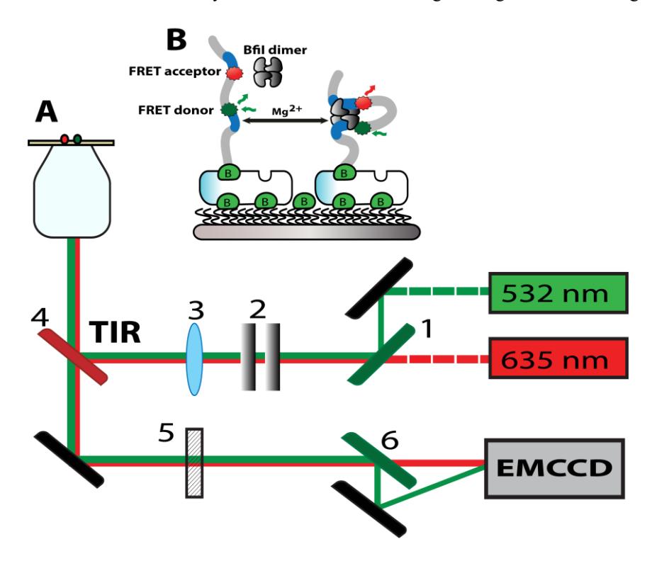
In this research we have studied interaction between DNA and BfiI restriction enzymes using single – molecule Forster Resonance Energy Transfer (smFRET) total internal reflection (TIR) microscopy. Biotinylated DNA molecules bearing two targets for BfiI and FRET pair dyes close to these targets were immobilized on a silanized and PEGylated (metoxy-PEG and biotin PEG mixture) glass surface via neutravidin. The protein induces formation of DNA loop: one BfiI site binds one target and second another, and therefore brings the FRET pair in close proximity (Fig. 1).
Our acquired smFRET signals have shown multiple FRET level lasting for several seconds. We observed that FRET efficiency and duration was affected by BfiI mutation, Cross-linking and Mg2+ ions/ ion strength.