
Unlike other biological fluids spinal fluid (SF) is much less often used to diagnostics of diseases. Anyway, despite the certain difficulties of SF sampling its biomedical research is the only reliable approach for diagnosing meningitis Traditional cytological, bacteriological, virological, and serological techniques are complex, expensive, require a lot of time and not always reliable. Raman spectroscopy (RS) undoubtedly is the most prospective way to SF researching because it is fast and non-invasive as well as provides reliable information about changes at the molecular level.
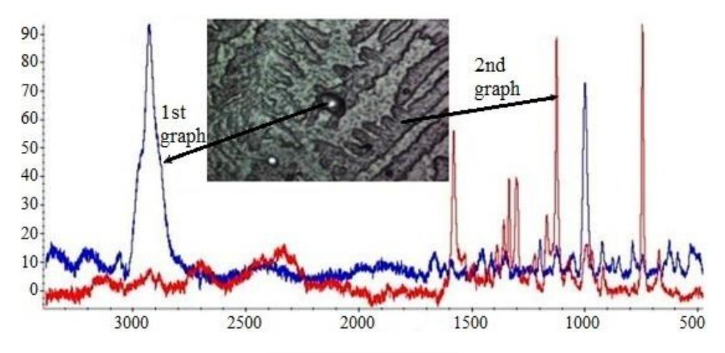
Researches to identify specific lines of bacteria which are the causative agents of meningitis were carried out. These lines can be used to efficiently detect and identify bacteria in the spinal fluid. Studies were carried out via Raman spectrometers Nicolet Almega XR and DXR2 with laser radiation at 532 nm and 785 nm wavelengths. Stokes components were registered in the range from 400 to 3100 cm-1. The measurements were carried out both by the traditional RS method and by the methods of surface-enhanced and resonant RS.
It is shown that the characteristic lines which can be used to detect bacteria are mainly due to the amide, protein, and DNA vibrations. For example, Staphylococcus aureus bacteria characteristic lines are associated with fluctuations of carotenoids (1528 and 1152 cm-1).
Thus, Raman spectroscopy of spinal fluid is quite useful way which could allow to quickly establish the cause of the meningitis in order to appoint the right treatment as soon as possible.