
It is important to determine radiation doses of patients in order to optimize the image quality contemporaneously with negative consequences of the radiological procedure. But there are a number of factors that cause uncertainties in the process of estimating doses to patients [1]. This paper is devoted to estimating the uncertainties that X-ray beam centration brings to the values of the absorbed doses in critical organs and tissues.
The thorax radiography in posterior-anterior projection was chosen as one of the most frequent X-ray examinations. In order to estimate the energy absorbed in organs and tissues the Monte-Carlo modelling of X-ray transport within the human body was performed. The reference voxel phantoms of an adult male and female were used as the models of human body [2]. All calculating were made for the following parameters: anode voltage 70 kV, 3 mm filtration of aluminum, voltage ripple 5%, the focus-film distance was 100 cm, the field size on the film was 30×40 cm2.
The reference centration (RC) was defined as the centration to the geometrical center of lungs. For the male phantom it corresponded to 40 cm and for the female phantom – 36.8 cm down from the top of the head. The cases of $\pm 5\%$RC, $\pm 10\%$RC, $\pm 15\%$RC and $\pm 20\%$RC to this centration were considered. The results of calculations were normalized by kerma-area product estimated for such examinations. Some of them are shown on the Fig. 1.
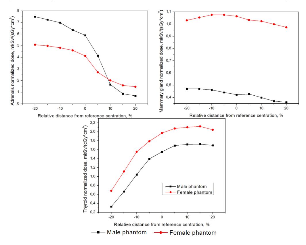
According to our research the position of X-ray beam causes large uncertainties (up to ten times) for organs that originally were out of the direct field of radiation or were partially in the field. For extended organs and tissues such as lungs, muscle or skin this changes are not so great (<20%) but nevertheless they should be taken into account for patient's dose assessment. Also it should be considered that doses to female organs practically in all cases are larger than doses to the same organs of male phantom. It can be explained by differences in their bodies' constitutions. Thus, all these findings will be taken into account by our team that develops the program for patients' doses assessment.