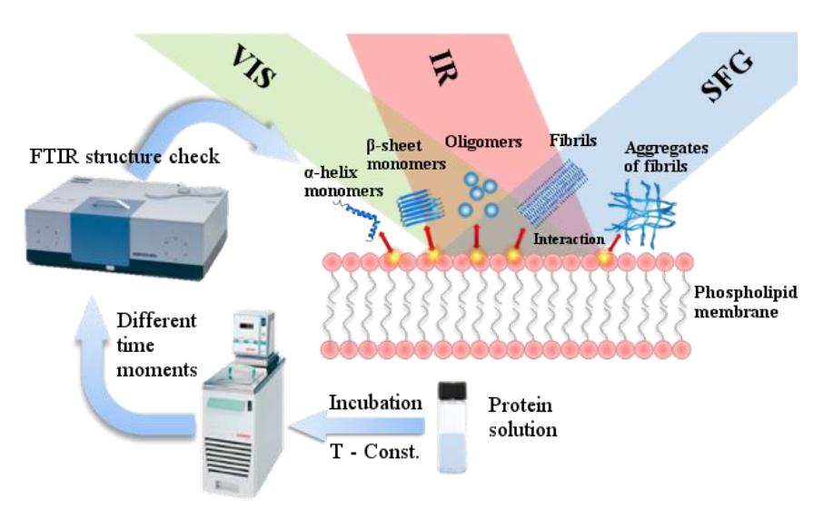
The formation of amyloid fibrils in cells and/or an intercellular net from primary disordered proteins is associated with more than 40 different clinical conditions such as Alzheimer's disease, Parkinson's disease, and type II diabetes. Amyloid $\beta$-protein (A$\beta$) is a normal product of cellular metabolism derived from the amyloid precursor protein (APP) by the successive action of the $\beta$- and $\gamma$-secretases [1]. Its elimination processes operating in parallel usually counterbalance this production of A$\beta$. When these production and elimination processes are disbalanced, proteins form different aggregates: small soluble oligomers and insoluble polymeric fibrils. All these aggregates are accumulating in brains and causing inflammatory processes, cell death, and tissue degradation.
The goal of our study is to investigate the aggregation process of protein on a molecular level. The most relevant question we aim to answer is how does the phospholipidic membrane of the cell influence the structure of the aggregates? It is believed that this interaction greatly affects the aggregation process therefore, it is important to understand how the molecular structure of aggregates changes at each stage of the formation due to the interaction with the membrane. To answer this questions a surface-sensitive spectroscopic method, which would allow probing molecules only in contact with lipid membrane is needed.
Vibrational sum frequency generation (VSFG) spectroscopy is a surface sensitive and molecular specific technique. It is widely recognized non-linear optical spectroscopic tool for studying the conformation and orientation of various adsorbed molecules at diverse interfaces. It is a second-order nonlinear optical process where two pulsed laser beams, one of fixed visible frequency and the other of tunable infrared frequency overlap spatially and temporally at an interface to generate a sum-frequency signal (schematics at fig. 1). The frequency of the generated VSFG signal is the sum of the two incident field frequencies and it is enhanced when the infrared frequency matches the vibrational frequency of the probed molecules. Moreover, VSFG can be used to unambiguously determine the chirality of the molecules. We applied SFG technique to study the structure of A$\beta$ fibrils. We measured the chiral SFG spectrum of A$\beta$ fibrils in Amide I band and NH vibration region. Our results confirmed NMR experiment [2] and showed that these fibrils are formed from parallel $\beta$-sheets.
In addition, we use Fourier transform IR (FTIR) spectroscopy to observe a protein aggregation process in the bulk solution. The repeat units in proteins give rise to nine characteristic IR absorption bands (amides A, B and I–VII) [3]. Amide I bands (1600-1700 cm-1) are the most prominent and sensitive vibrational bands of the protein backbone, and they relate to protein secondary structural components.