
Fluorescence microscopy is a well-known imaging technique that shows specific protein distribution in cells. In recent years, combination of fluorescence microscopy and Raman spectroscopy is also used to provide both finding and identification of different organic molecules. However, the most currently available fluorescent organic dyes are characterized by relatively weak emission, rapid photo-bleaching and tendency to hide Raman signal. Alternatively, metallic nanoparticles have also been extensively applied for cellular imaging and biomedical diagnostics. For example, inorganic metallic nanocrystals known as quantum dots (QDs) are often used as fluorescence labeling agents for biological cells and tissues. QDs offer significant advantages over conventional organic dyes such as brighter fluorescence and higher resistance to photo-bleaching that make them suitable for ultra-sensitive imaging [1]. What is more nanoparticles of noble metals demonstrate activity in surface-enhanced Raman scattering (SERS) spectroscopy which opens prospects of a detection sensitivity of 16 orders of magnitude higher than that of ordinary Raman spectroscopy [2]. As a result, recovery of Raman signal from fluorescence background is observed. In this paper, we report on visualization and SERS-detection of organic dye R6G that is mixed with gold colloids and adsorbed on the SERS-active substrate based on the silvered porous silicon.
SERS-active substrates were formed by electrochemical etching of highly-doped n-Si wafers in HF-based solution that resulted in fabrication of mesoporous silicon layer of 5 μm thickness and following electroless deposition of silver nanoparticles of 10 – 150 nm diameters. The concentration of gold nanoparticles in colloid was $4\times10^{-4}$ mol per liter of water while their diameters varied from 10 to 20 nm. An analyte for the SERS and fluorescence measurements was prepared by mixing water solution of $10^{-9}$ M R6G and gold colloid in 1 : 9 volume ratio. As a result, total concentration of R6G was $1\times10^{-10}$ M. This mixture was drop-deposited on the porous silicon coated with silver nanoparticles and dried in the air. Optical microscope equipped with filter provided transmission in the range of 330 – 385 nm was used for the fluorescence imaging. SERS measurements were performed using 532 nm laser.
The analyte drop formed so-called "coffee ring" on the substrate after complete drying because of agglomeration of gold nanoparticles. Fig. 1 shows fluorescence image and SERS spectrum of R6G drop-deposited on the silvered porous silicon from its mixture with gold colloid.
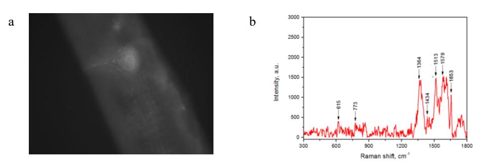
Following Fig. 1, we can observe the highest fluorescence in the area of "coffee ring". Despite the SERS spectrum (Fig. 1, b) is a little bit noisy, typical bands of R6G ( 615 cm-1; 773 cm-1; 1364 cm-1; 1434 cm-1; 1513 cm-1; 1579 cm-1; 1653 cm-1) are distinguished. The ability to register SERS spectrum is caused by the rough surface of the silvered porous silicon that provides absorption between 370 nm and 800 nm due to an arrangement of surface plasmon resonance in this range [3]. The most intensive absorption is in near UV region. That is why, SERS spectrum is not completely hidden in fluorescence background.
Summarizing, the combination of fluorescence and SERS techniques allowed us to find and identify organic dye R6G drop-deposited from the solution of $1\times10^{-10}$ M. This opens an opportunity of ultrasensitive bioimaging and molecules identification.