
Self-assembled monolayers (SAMs) from organethiol molecules adsorbed on noble metals are widely used to create interfaces with desirable properties. SAMs with positive charge bearing terminal group have attracted growing attention in the last few decades for few reasons. Such SAMs have electrostatic ability to attract inorganic anions and peptides from the solution phase. In addition, monolayers bearing positively charged terminal group have been used for sensing the anions and for the fabrication of bioelectronics devices [1, 2].
Surface-enhanced Raman spectroscopy (SERS) is one of the most sensitive vibrational spectroscopic method for probing the bonding, structure, and orientation of adsorbed molecules at certain nanostructured metals (mostly Au, Ag, and Cu). On the other hand, the limited number of SERS-active substrates and requirement to use roughened surfaces restricts the applicability of this method for analysis of technologically important surfaces. Consequently, Tian et al. approached a novel SERS technique named "shell-isolated nanoparticle-enhanced Raman spectroscopy" (SHINERS). The method is based on enhancement of Raman signal by strong electromagnetic field provided by gold core nanoparticles surrounded by a few nanometre thick inert silica shell (Au@SiO2) [3].
The present work focused on in situ SHINERS study of positive charge bearing SAM formed from N-(6-mercapto)hexylpyridinium (MHP) at smooth Au electrode in aqueous NaClO4 solutions. SHINERS was used to probe the electrochemical potential effect on bonding with surface, ion-pairing, and molecular structure of MHP.
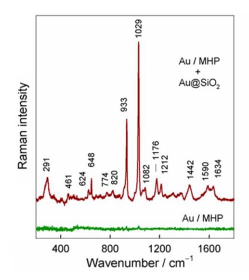
During this study, it was found that metal-adsorbate vibrational mode $v$(Au-S) was detected in the SHINERS spectra at 282-305 cm-1 (Fig. 1). Near linear potential-induced blue shift of this mode was revealed as electrode potential was tuned to more positive values. Spectroscopic analysis showed formation of ion-pairs between the positively charged pyridinium group of MHP and $ClO_4^-$ anions in the solution phase.
SHINERS is emerging recently as a novel technique that uses shell-isolated nanoparticles to improve the applicability of SERS. SHINERS overcomes the long-term limitation of SERS because it allows SERS measurements on various target systems that were inaccessible previously because of the material and surface morphology issues. Moreover, SHINERS can be used in many complicated environments such as in aqueous solutions and biological systems.