Recently 3D printing technologies have received a lot of attention in the areas of science and industry. Differently from traditional processes of manufacturing, 3D printing allows fabrication of arbitrary geometry objects, using computer aided design and spatial formation of the structures layer by layer. It can be applied in various fields. One of them is medicine, where 3D printed objects have already made a great influence in such areas as orthopedics [1], face and skull reconstruction [2], plastic surgeries [3], teeth and mouth operations [4], etc.
Cells can be seeded into 3D printer-formed structures and artificial tissue or organ can be grown and implanted into human body. Currently, there is a lack of elastomeric photostructurable resins that would fit for tissue engineering requirements for scaffolds: non-degradable, but also biocompatible; permeable to low molecular weight molecules, gases and nutrients, avoiding waste accumulation; pore size should be appropriate for cell to cell interaction and migration [5]. However, optimizing biocompatibility of existing commercial resin would make a great influence. Application of new materials would open the door for new tissue engineering possibilities, enabling fabrication of the scaffolds with different mechanical, physical, biochemical and electrical properties that are needed for different types of tissues [6].
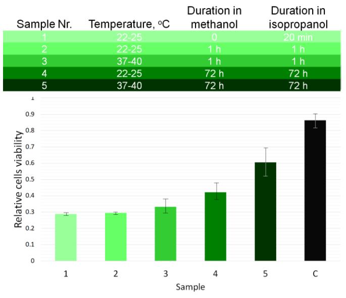
In this work the principles of fabrication of 3D scaffolds via stereolithography and scaffolds' biocompalibilty are investigated. In this research the requirements of reproducing the original environment of cells in such bioscaffolds are used in order to create and test mechanically flexible microporous structures. The scaffolds were fabricated using the tabletop 3D printer Autodesk Ember. Commercial photoresin Formlabs Flexible was used for this task. Optimization steps presented in this paper allowed to increase the biocompabaility of the scaffolds by 48% in comparison to unoptimized ones.
| Sample Nr. | Temperature, °C | Duration in methanol | Duration in isopropanol |
|---|---|---|---|
| 1 | 22-25 | 0 | 20 min |
| 2 | 22-25 | 1 h | 1 h |
| 3 | 37-40 | 1 h | 1 h |
| 4 | 22-25 | 72 h | 72 h |
| 5 | 37-40 | 72 h | 72 h |