
Magnetic resonance imaging today is a powerful method of studying the internal structures of the human body. Its wide application it received due to diagnostic woes and virtually no harmful effect on the patient. In connection with a wide range of applications, the actual tasks that physicists and engineers are working on are improving the accuracy of diagnostics, reducing measurement time, obtaining data on biophysical and biochemical processes. The characteristics of the magnetic fields used have a significant effect on the solution of the above problems, among which it is important to note the magnitude of the constant magnetic field, the parameters of spatial instability or inhomogeneity. The solution of the problem of compensating the instability of magnetic fields remains relevant in the tomography. An example of such studies is the research on the correction of distortions caused by the heterogeneity of the magnetic fields [1].
The work is devoted to the development of a technique for reconstruction of images from partially incomplete data of measured signals. This technique is useful in solving problems of reducing the time of measurement. During the measurements, the patient is in the measuring area of the tomograph and must be as much as possible immobile. His movement leads to the appearance of artifacts. In addition, some of the measured data can be distorted by electromagnetic processes in the receiving system. These distortions can be cased by random significant amplitude fluctuations of the signal. In this work, we simulate the recording of signals based on the processing of a real tomographical image (Fig.1,a). For simulation we considered the free-induction decay signal, Eq. (1) for constructing tomograms weighted by spine density $\rho(x, y)$ in gradient k-space $(k_x,k_y)$.
$$S(k_{xl},k_{ym}) = \widehat{S} \sum_{a,b} \rho(x_a,y_b) \cdot e^{i(k_{xl}x_a + k_{ym}y_b)}, \tag{1}$$where $\widehat{S}$ is a constant, $k_{x,y} = \gamma G_{x,y} \tau_{1,2}$ ($\gamma$ - hydrogen gyromagnetic ratio; $G_{x,y}$ - magnetic fields transverse gradients; $\tau_{1,2}$ - MR pulse sequences delay time characteristics).
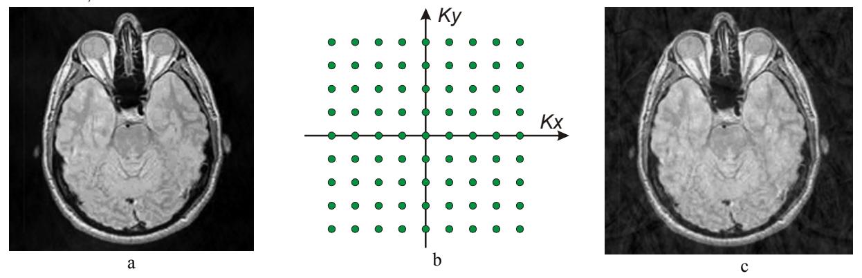
To carry out the reconstruction a complete set of signals is necessary in the corresponding to the space-frequency two-dimensional spectrum (K-space, Fig.1,b). We considered the array of the signals with fullness from 50 to 90 percent with 10 percent increments. The reconstructed image for completeness of 90 percent is shown in Fig.1,c. Missing data on the gradient grid in K-space were calculated by interpolation over available nearest points [2]. This image can be measured in a real tomograph for 10 percent of the time faster, and this is a few minutes. During this time, it is possible, that the patient will not make a significant movement, then the quality benefits will be even more obvious. It should be noted that such measurements can be carried out when it is known in advance that the quality requirements can be reduced. A second option for applying the method can be the processing of already available data that have the above described inaccuracies. The third application of the proposed approach is the development or calibration of tomographical systems.