
Sodium dodecyl sulfate (SDS) is one of the most commonly used detergents in protein chemistry. It has shown benefits for sample preparation, including protein solubilization and denaturation. SDS is typically used at concentrations between 0.1 % and 2%, but application of 4% SDS was also reported [1]. However, application of SDS in sample preparation for liquid chromatographic – mass spectrometric (LC-MS) analysis is limited, because SDS is recognized to cause significant chromatographic peak broadening in reversed phase liquid chromatography and signal suppression in electrospray ionization mass spectrometry. Given the advantages of using SDS for protein sample preparation, detergent removal prior to LC-MS analysis is often performed.
It was clearly reported that threshold tolerance of SDS in LC-MS experiments is 0.01 % [2]. Several different strategies can be applied to lower SDS concentration below this level. In order to compare the effectiveness of different SDS removal methods for a given protein sample, a simple and accurate SDS determination method is required. Mukerjee suggested photometric SDS determination method based on organic dye methylene blue and dodecyl sulfate ion pair extraction from aqueous solution to chloroform [3]. This strategy was reported being compatible with a number of biochemical reagents, nucleic acids and proteins [4]. However, our results show that high concentration of protein prevents methylene blue extraction to organic phase (see Fig. 1).
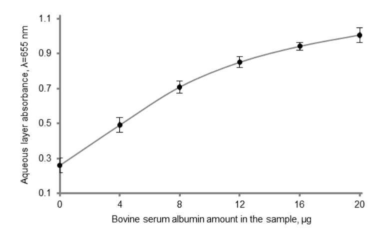
In this work we present a simple modification of Mukerjee's method, which enables SDS concentration measurement in samples initially containing 1.8 mg/ml bovine serum albumin. Our modified method includes cold acetone protein precipitation procedure, which is applied to both protein samples and calibration curve SDS standards before methylene blue – dodecyl sulfate extraction. Protein removal prevents precipitate formation between aqueous and chloroform layers, thus ensuring efficient ion pair extraction. Moreover, we measured absorbance of the aqueous layer instead of measuring absorbance of organic phase. This is in contrast to reported procedures, but aqueous layer is compatible with disposable polystyrene cuvettes while chloroform requires cuvettes made of glass. The method reported is accurate (relative error < 7 %) and relative standard deviation is less than 5 % (n = 8). The concentrations and the volumes of reagents were optimized so that the method is suitable for monitoring of SDS at concentrations between 0.002% and 0.025%. The method was successfully used for comparison of different SDS removal strategies.