
Introduction: Brachial plexus anatomical variations is important to anesthesiologist who administer regional anesthetic blocs1. The cognition of these variations helps in clinical practice.
Aim: The goal of this study was to scan posterior triangles of the neck by ultrasound and identify the relationship between brachial plexus and scalene muscles, compare results with scientific literature data and evaluate anatomical variations prevalence depending on age and sex.
Materials and methods: 100 participants (38 men, 62 women) were examined by ultrasound. Scanned 200 brachial plexus altogether. All anatomical variations have been classified. Age and sex were noted and relation between anatomical variations were checked.
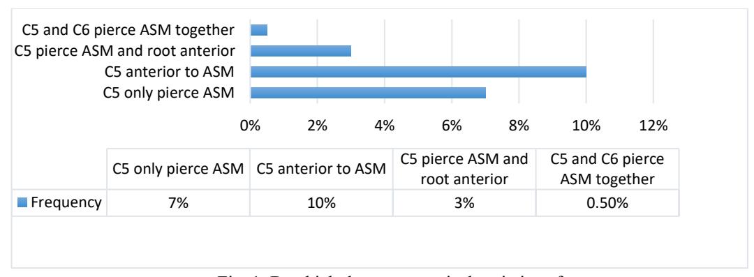
Results: The mean of age of participants were 33.63 (SD 12.4) years. Only 3.5% (n=200) of brachial plexus were not identified, (bilateral 6%). C5 and C6 roots tracked the interscalene gap (ISG) 76% of all cases, bilateral 63% and it is the commonly described anatomical position. C5 root tracked anterior to anterior scalene muscle (ASM) 10% (bilateral 4%), C5 only pierce ASM 7% (bilateral 2%), C5 pierce ASM and root anterior 3% (bilateral 2%), C5 and C6 pierce ASM together 0.5% (bilateral 0%) of all cases (Fig. 1.). No statistically significant correlation was found between sex, age and distribution of anatomical variations. However, some findings showed that older participants had the same anatomical variants bilateral more often (p=0.048).
Conclusion: Ultrasound guided examination of posterior triangles of the neck is relatively onefold, because the structures are superficial and easily visualized. Anatomical variations described as normal have been reported more often (76%) than published in literature and abnormal cases have been found less (0.5-20.5%) respectively1-3. There was no statistically significant value between sex, age and distribution of anatomical variations.