
Biofilms are implicated in approximately 80% of all chronic infections. Their prevalence in conditions such as chronic wounds, catheter-associated infections and pneumonia underscores the necessity for effective detection methods to manage these infections properly [1]-[2]. Electrochemical techniques can provide quick results, often within minutes to hours, compared to the days required by traditional culture methods [3]. Identifying biofilm presence can inform treatment decisions and improve patient outcomes [4]. This work seeks to determine the electrogenic characteristics of bacterial biofilms from Gram-positive species S. haemolyticus, E. faecalis and Gram-negative species P.aeruginosa, S. maltophilia.
To monitor biofilm growth, we used open circuit potential (OCP), chronoamperometry and optical density (OD) measurements. A carbon cloth (CC) and Ag/AgCl 3M KCl were used as working and reference electrodes, respectively, as presented in the schematic illustration (Fig. 1). Depending on the bacteria, the OCP shifted from 100-50 mV to –200 - –300 mV in a bacterial solution, indicating that bioelectrochemical coupling had occurred. We observed the negative potential shifts by mediated electron transfer (MET), which connects bacterial metabolism to the electrode surface. Amperometry further confirmed the bacteria’s ability to transfer electrons through MET and direct electron transfer (DET) mechanisms. In agreement with OCP and OD results, the current-time dependence highlights the extracellular electron transfer (EET) mechanism across different stages of biofilm development and bacterial growth, including the lag, log and stationary phases.
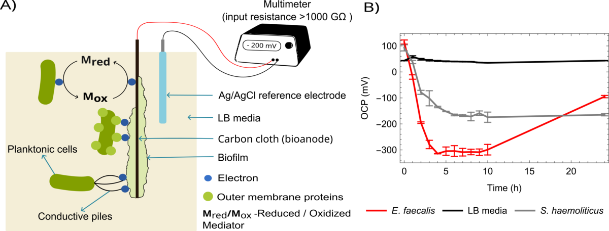
This study shows that E. faecalis had the most negative OCP (-303 mV) and the highest current density (435 nA/cm\(^{2}\)), whereas S. haemolyticus demonstrated the lowest electroactivity with an OCP of -165 mV and a current density of 120 nA/cm\(^{2}\). Gram-negative bacteria exhibited stable OCP and steady currents, reflecting consistent bioelectrochemical activity. These results highlight the potential of electrochemical methods for rapid detection and analysis of biofilm-related infections.