Plastic scintillating optical fibers have gained attention in high energy particle tracking detectors due to their flexibility, ease of fabrication, low cost, good light output, good timing and insensitivity to magnetic fields. They are employed in major experiments such as the Large Hadron Collider beauty (LHCb) experiment, where they are the central component of SciFi detector [1].
The aim of this study is to investigate distribution of various spectral properties across the cross-section of plastic scintillating optical fibers. Assessing the homogeneity of these fibers is important for ensuring uniform signal formation and accurate particle detection needed for tracking detectors application.
Two different plastic scintillating optical fibers (Kuraray) were analyzed: 78MJ and 3HF, with luminescence peaks at 450 nm and 520 nm, respectively. They are 250 μm in diameter, with structure consisting of core with activator and spectral shifter luminophores, inner polymethyl-methacrylate (PMMA) cladding and outer fluorinated polymer cladding [2].
The uniformity of samples was analyzed by investigating the spatial distribution of different photoluminescence parameters. The investigation was performed using the WITec Alpha 300S scanning confocal microscopy system coupled with a 405 nm excitation laser. Measurements were performed across the entire fiber cross-section and separately on the core center, side and cladding, using objectives with different magnifications and numerical apertures.
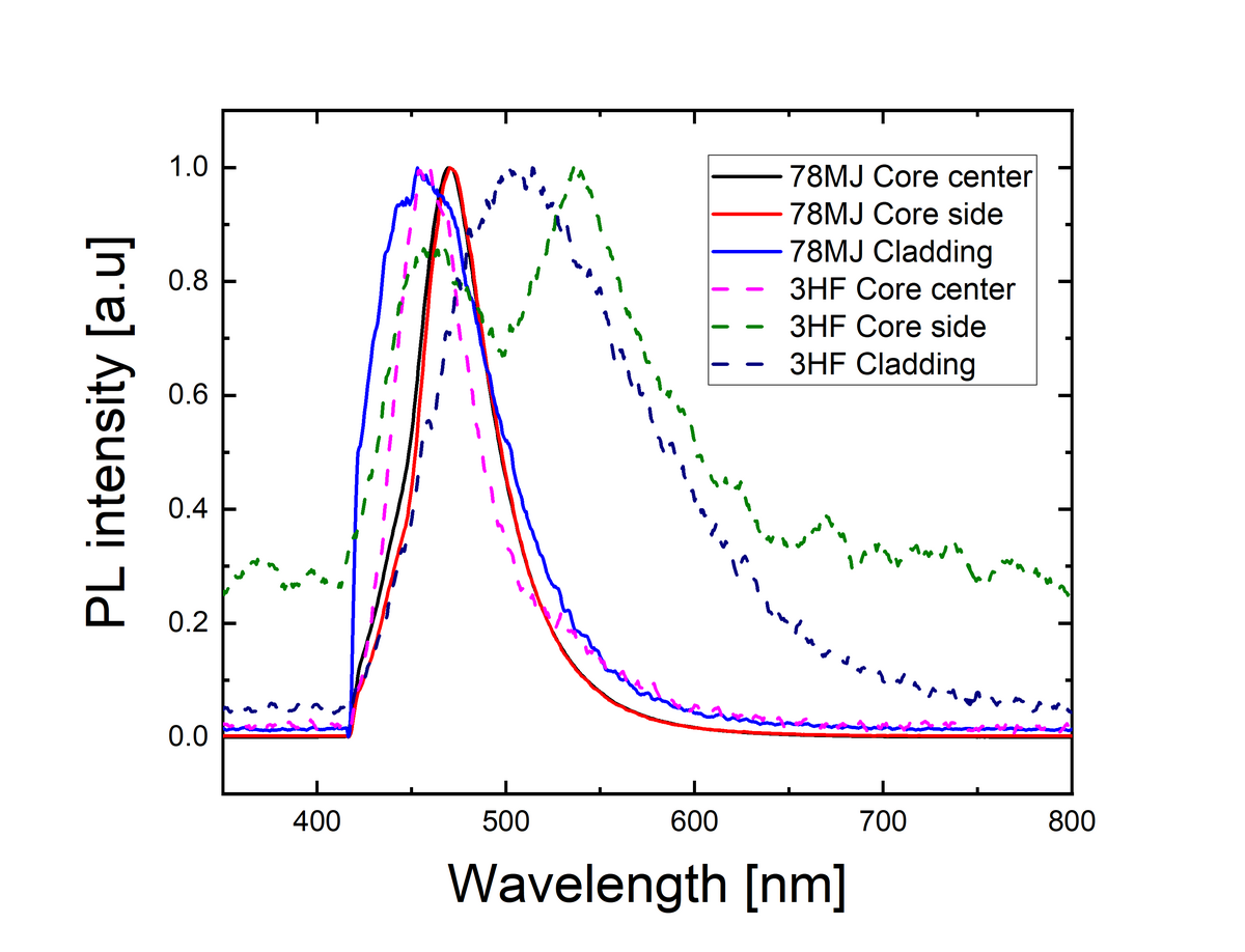
The measurements showed higher uniformity in 78MJ optical fiber with both core center and sides scintillating at 470 nm while cladding showed 450 nm peaks. In contrast, the 3HF fiber displayed greater spectral variation, with the core center scintillating at 460 nm and sides showing second peak at 540 nm. Also more localized peaks of 470, 530 and 450 nm were detected. 3HF fiber cladding showed peak at 500 nm. These results will be used for further research and serve as a reference for comparison with radiation-exposed plastic optical fibers.