$ AGGREGATES USING ATOMIC FORCE MICROSCOPY} The adsorption of molecules or nanostructures on solid surfaces is one of the fundamental processes for the development of molecular-based nanodevices. Atomic force microscopy (AFM) is a microscopic technique with extremely high lateral resolution for surface imaging. AFM imaging technique was used to reveal TPPS\({_4}\) aggregate geometric structures, ranging from roughly spherical to highly irregular structures with dimensions in the nanometer scale.
Here we focused our investigation on the adsorption of the TPPS\({_4}\) aggregates deposited onto hydrophilic mica substrate. 5,10,15,20-Tetrakis(4-sulfonatophenyl) porphyrin (TPPS\({_4}\)) solutions, containing aggregates, were deposited onto mica substrates, dried and/or spin-coated, and then imaged using AFM.
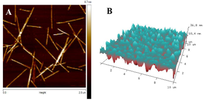
Images of the aggregates deposited on mica substrate are presented in Figure 1> TPPS\({_4}\) aggregates consisting of individual rods and bundles of rods (A - pH=3) and complex layers (B - pH=7) are seen in the image. The single aggregate rods have a remarkably uniform height, and roads appear to be the basis buildings blocks for longer bundled aggregates [1].
Our prior spectroscopic and AFM investigations [2] showed different aggregation patterns are observed when samples are prepared using TPPS\({_4}\) solutions of different pH values.
Studies on the spatial properties of TPPS\({_4}\) aggregates formed in solutions allowed us to visualize result of aggregation processes in different pH environments. Interestingly the formation of aggregates in neutral media was observed in spectral measurements but was not observed by AFM. Such structures offer significant potential for the development and fabrication of diverse nanoscale optoelectronic devices.