Optical coherence tomography (OCT) is an interferometric imaging technique commonly used for visualizing various ex vivo and in vivo biomedical samples. The axial resolution of an OCT system is mainly dictated by the spectral width (and consequently the coherence length) of the light source and lateral resolution by the numerical aperture of the objective lens. Full-field (FF-) OCT can achieve resolution better than 1 μm because it allows use of high-NA objectives and broadband light sources [1,2].
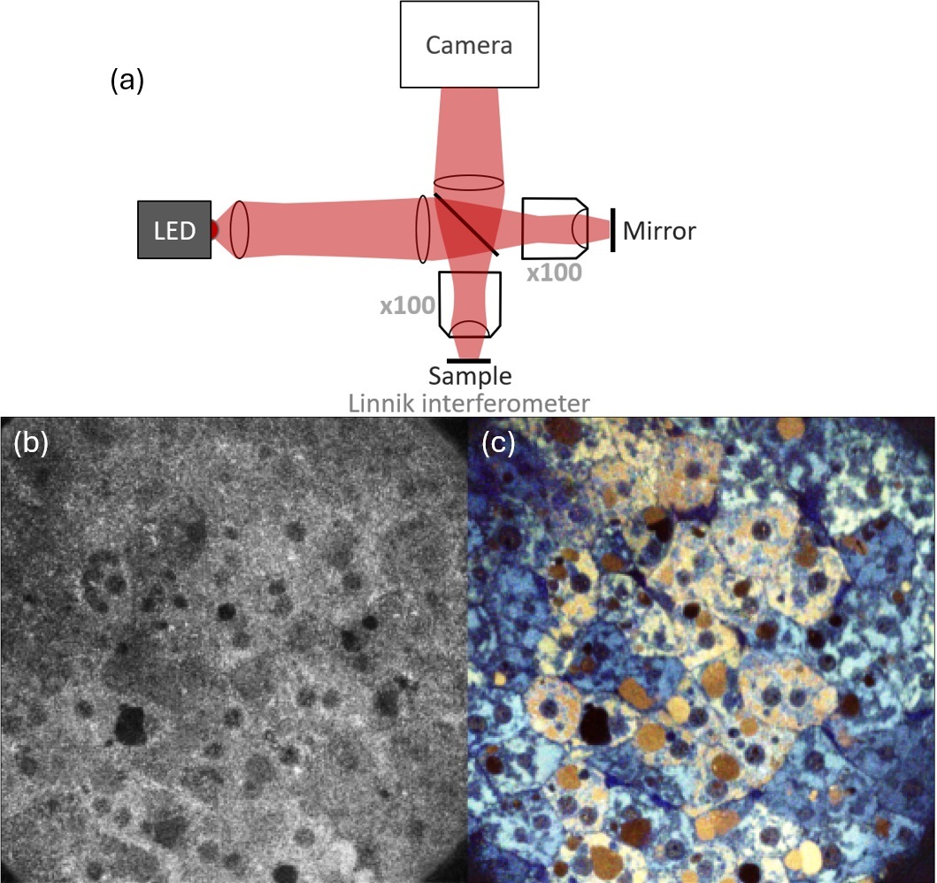
Fig. 1. Principle schematics of high-resolution FF-OCT system (a), comparison between regular (b) and dynamic FF-OCT (c) images of mouse liver.
High-resolution FF-OCT system, shown in Figure. 1.a, can image deep in tissue, featuring Linnik interferometer with two identical high NA immersion objectives, a powerful spatially incoherent white light source and a specialized camera. The system also includes a piezo actuator to enable phase shifting that is necessary to extract OCT images.
Figure 1.b shows a regular FF-OCT image of mouse liver acquired through phase-shifting, wheras Figure 1.c shows dynamic FF-OCT image derived by analyzing vibrations in the acquired interferometric images. As seen in these images, the dynamic FF-OCT provides more detailed images, with clearer cell walls and distinct structures of hepatic tissue. This imaging technique enables the visualization of different cells without the need for specific preparation methods, such as dyes or other modifications commonly required in other imaging techniques.
[1] Dubois, A., Grieve, K., Moneron, G., Lecaque, R., Vabre, L., & Boccara, C. (2004). Ultrahigh-resolution full-field optical coherence tomography. Applied Optics, 43(14), 2874. https://doi.org/10.1364/ao.43.002874
[2] Monfort, T., Azzollini, S., Brogard, J., Clémençon, M., Slembrouck-Brec, A., Forster, V., Picaud, S., Goureau, O., Reichman, S., Thouvenin, O., & Grieve, K. (2023). Dynamic full-field optical coherence tomography module adapted to commercial microscopes allows longitudinal in vitro cell culture study. Communications Biology, 6(1). https://doi.org/10.1038/s42003-023-05378-w
[3] Trečiokaitė, A., Adomavičius, K., & Auksorius, E. (2024). Time-domain full-field optical coherence tomography with a digital defocus correction. Optics Letters, 49(10), 2605. https://doi.org/10.1364/ol.520911