Numerous kinds of nanoparticles are getting increasingly popular in biomedicine. Different nanoparticles are designed considering their desired use, function, modifications. Upconverting nanophosphors (UCNPs) have the ability to absorb two or more lower energy photons to yield a single higher energy photon. This is especially useful when using them on tissues below surface. Bearing in mind an undesirably high tissue absorption in UV range, which is used to excite other nanoparticles and tissue autofluorescence under UV excitation UCNPs can be irradiated in up to 1 cm depth and prevent tissue luminescence. Using various combinations of dopant ions allows for spectral nanoparticle tuning, making it even more versatile in biomedical applications [1]. The purpose of this paper is to investigate the UCNP's ability to be absorbed into cells and used as an imaging marker, possible cytotoxicity and colloidal stability.
For these measurements we used NaYF4:Yb3+,Er3+ nanoparticles modified with F127 surface coating polymer. Nanoparticle colloidal stability was measured in relation to luminescence intensity in a solution. Different acidity solutions were compared, as well as different cell incubation mediums. Our results show increased colloidal stability in solutions set at pH 7 and 7.4 and low intensity luminescence relating to low colloidal stability in pH 6 and pH 8 phosphate buffer solutions. While comparing different cell mediums we have found that Fetal Bovine Serum (FBS) presence in Dulbecco Modified Eagle's Medium (Gibco, USA) solution extends UCNP solubility compared to DMEM solution without FBS and deionized water. Transparent cell mediums with FBS were also used to increase luminescence intensity by avoiding solution absorption (DMEM and OptiMEM), both of which allowed for higher colloidal stability.
Cell cytotoxicity evaluation was carried out using standard XTT assay. MCF 7, MDA MB 231 and MCF 10A cell lines were used. Cells were incubated using UCNP concentration, equal to that used in cell imaging, along with two and five times increased concentration. After up to 24 hour exposure to UCNPs in various concentrations cell viability dropped to no less than 80 percent in all cell lines, as evaluated by XTT assay.
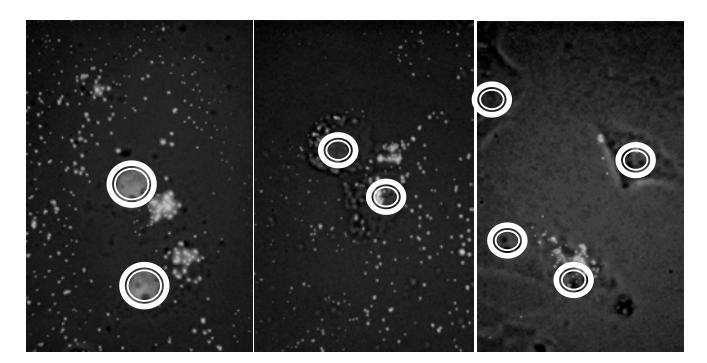
Cell imaging was carried out using a custom modified Nicon Eclipse E400 microscope equipped with 980 nm continuous wave operation diode laser with emission power of 877 mW for nanoparticle excitation. Cell nuclei were stained with 4',6-diamidino-2-phenylindole DNA binding dye. Cells were incubated with UCNP solution for 1, 3,6 and 24 hours. Cell images were processed using ImageJ software (Fig. 1) (NIH, USA).