This research is directed towards the multivariate analysis of native fluorescence (also known as auto-fluorescence) of human intervertebral disc samples for medical classification purposes.
During operations of intervertebral disc hernia in Clinics of Neurology and Neurosurgery pieces of intervertebral disc tissue were removed as a part of conventional procedure. Autofluorescence of these samples were excited in-vitro with 355 nm radiation (microlaser STA-01-TH, Standa, ltd.) and registered with spectrometer OceanOptics USB2000 through a system of fiber optics. The specimens were classified to three medical groups in accordance with localization of tissue (D - specimen from inner part of a disc, S - free sequester, P – herniation from subligamentous space). In this investigation 527 spectra (group D – 38%, group P-41%, group S – 21%) were investigated.
Spectral data reduction was carried out by means of multivariate curve resolution methods (such as multivariate curve resolution-alternating least squares and principal component analysis) utilizing 11 component model, which gave the highest performance in the previous investigation (unpublished data). The performance of group identification was performed pairwise (D vs. P, D vs. S, P vs. S, and S vs. all remaining groups, i.e., others, P vs. others, D vs. others.) applying receiver operating characteristic (ROC) method.
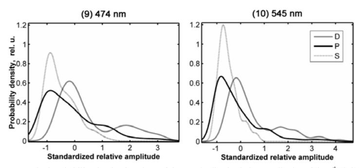
Results. Component nr. 11 was considered to be a noise due to extremely low amplitude. The best performance to discriminate groups was achieved using the long-wave side components. Despite the fact that the amplitudes of components are overlapped the tendencies to separate spectra can be noticed in the graphical representation of results (Fig.1). The accuracy higher than the minimum requirement for medical tests (i.e. area under the ROC curve, the AUC, is greater than 0.75) was achieved.