Biomimetic lipid membranes are widely used in vitro systems to study membrane lipid or protein-protein interactions. Many effects of membrane composition, phase behavior and transport can only be studied by in vitro reconstitution. Combination of self-assembled monolayers (SAMs) on solid supports and the direct nano-patterning technique known as dip-pen nanolithography (DPN) allow controlling the composition and geometry of the biomimetic lipid assembly at the nano scale.
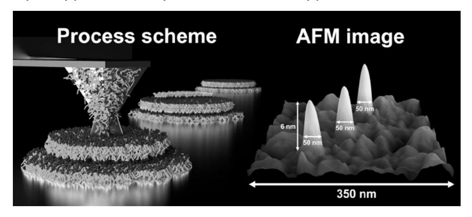
The aim of this study was to explore the possibilities to combine lipid DPN [1] with micro-patterned SAMs. To characterize the lipid ink (1, 2-dioleoyl-sn-glycero-3-phosphocholine) behavior, two homogeneous SAMs were used: hydrophobic eicosanthiol (C20) and hydrophilic tri(ethylene glycol) - terminated thiol (EG3). To increase the reproducibility, an ink-jet inking system was used to equalize the lipid ink amount on the tip. Furthermore, all experiments were done in a controlled humidity chamber at 50% RH and 25 °C. Lipid patterns were investigated using imaging ellipsometry and atomic force microscopy (AFM). Combination of these techniques allowed us to perform a comparative analysis that revealed sharp contrasts in the lipid mass flow rate and phase behavior on C20 and EG3 SAMs, respectively. We calculated that the transfer rate on the EG3 SAM was about 10 times greater (depends on the contact time) than on C20. However, due to the surface hydrophobicity the lipid phase behavior was completely different. Lipid patterns on the EG3 SAM formed a monolayer less than 1 nm in height. In contrast, on the hydrophobic C20 SAM, lipids molded into dot-like multilayer stacks over 200 nm in height. A decrease of the contact time allowed forming lipid dots less than 50 nm in diameter and 6 nm in height. Lipid DPN on the pre-structured surfaces with areas of hydrophobic and hydrophilic SAMs revealed that lipids preferably spread along hydrophilic areas, while hydrophobic ones acted as a barrier for spreading.
Based on these observations we were able to guide DPN lipid spreading and form lipid domains of pre-defined shape and size. These results promote further development of micro and nano-patterned lipid domains for creation of a simplified biophysical model of the complex cell membrane assemblies for biophysical studies.