
Ovarian cancer accounts for about 3% of cancers among women, but it causes more deaths than any other cancers of female reproductive system [1]. Due to the lack of specific symptoms, about 70% of ovarian cancer cases are diagnosed in advanced stages, when five-year survival rate is less than 40% [2] and standard treatment (cytoreductive surgery and chemotherapy) is often insufficient. It is known that ovarian cancer frequently tends to develop resistance to chemotherapy. Novel models and biomarkers are required for extensive research of drug resistance mechanisms.
The aim of this study was to identify the changes in the expression of drug resistance-related genes (PCNA [3], ASS [3], FN1 [3], ATPB7 [4], ATPB11 [5], NOTCH3 [6], MMP3 [7], NACC1 [8], ABCC1 [9], ABCC5 [9], ABCC10 [9]) in drug-resistant sublines of human epithelial ovarian cancer cell line A2780, developed after treatment of the original cell line with the most commonly used medicaments: paclitaxel, carboplatin, cisplatin and doxorubicin.
Cell line A2780 was chosen as a model for investigation of drug resistance in ovarian cancer. Cells were maintained as monolayer in medium (RPMI) supplemented with 10% fetal bovine serum, 2 pmol/L L-glutamine and antibiotics (streptomycin and penicillin) at 37°C in a 5% CO2 atmosphere.
Cell line was treated with different chemotherapeutic agents. The doses of the drugs were increased constantly with respect to resistance development. Different sublines were developed: A2780P, A2780Cis, A2780C, A2780D, treated respectively with paclitaxel (up to 900 ng/mL), cisplatin (up to 50 μg/mL), carboplatin (up to 15 μg/mL) and doxorubicin (up to 5 μg/mL). Flow cytometry was used to assess the cancer stem cell phenotype by staining cells with monoclonal antibodies against surface proteins CD24, CD44 and ESA.
Treated cells sublines were also analyzed for gene expression changes. After total mRNA extraction and cDNA synthesis, real time PCR was performed to evaluate the changes in expression of PCNA, ASS, FN1, ATPB7, ATPB11, NOTCH3, MMP3, NACCI, ABCC1, ABCC5, ABCC10 genes.
The development of drug resistance was confirmed by the increase of the median lethal dose (LD50) in all treated sublines: 2,7-fold increase in A2780P, 1,75-fold increase in A2780Cis, 3,1-fold increase in A2780C and more than 5000-fold increase in A2780D.
The most pronounced changes in cell morphology were observed in sublines treated with cisplatin and doxorubicin. In addition, the major increase of stemness marker expression was also observed in these particular sublines. The expression of ESA increased from 20% in original cell line to 100% in both resistant sublines. The expression of CD44 increased from 0% in original cell line to 85% in A2780D and 95% in A2780Cis.
The major changes in gene expression were observed in FN1, NOTCH3 and MMP3 genes, especially in A2780Cis and A2780D sublines (Fig.1.). Proteins encoded by these genes are responsible for cell adhesion, migration, growth, differentiation, and integration in the tissue. Changes were also detected in PCNA, ATPB7, ABCC1, ABCC5, ABCC10 genes. No significant changes were noted in ASS and ATPB11 genes.
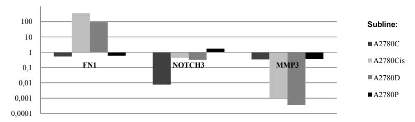
The results imply that A2780 cell line is a suitable model for investigation of drug resistance in ovarian cancer. Significant increase in FN1 gene and decrease in MMP3 gene, together with increase in stemness marker expression and morphological change are characteristic for doxorubicin- and cisplatin-resistant sublines. It is necessary to further investigate these features in ovarian tumor specimens for confirmation and translation of the results into clinical practice.