
Magnetic nanoparticles are a class of nanoparticle which can be manipulated using magnetic field. Ferrous or ferric oxide is the main constituent of magnetic particles, although metals such as cobalt and nickel are used in other fields of application. Magnetic nanoparticles, with their unique magnetic properties and controllable sizes, are being actively investigated as the next generation of magnetic resonance imaging contrast agents. Furthermore they have important potential for hyperthermia therapy, anticancer drug delivery and magnetic separation [1, 2].
The aim of our work was to investigate the accumulation of Fe3O4 nanoparticles in mouse embryonic fibroblasts (NIH3T3) and their effect on proliferation and viability of cells.
Fe3O4 nanoparticles were synthesized according to previously reported procedure [3]. Atomic force microscopy measurements showed that height of Fe3O4 particles varies from 10 nm to 40 nm, width – from 50 nm to 200 nm. Hydrodynamic size measurements revealed that the diameter of particles in solution is approximately 50 nm.
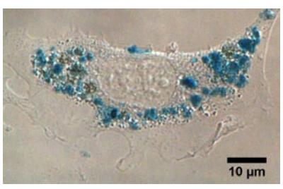
Cellular uptake was investigated incubating with 65 ng/ml of Fe3O4. For uptake studies of Fe3O4 nanoparticles in NIH3T3 cells we used Prussian Blue staining reagent, which reacts with the iron producing a blue color. The accumulation was observed using bright-field microscope equipped with digital color camera. Fe3O4 nanoparticles accumulated in cells heterogeneously: in some cells Fe3O4 nanoparticles were localized in large vesicles spread through whole cytosol, while in other cells vesicles were localized only in the perinuclear region (Fig. 1).
Toxicity of Fe3O4 nanoparticles was investigated using XTT cell viability assay. The viability of NIH3T3 cells remains approximately 98% within 3-24 h of incubation, however a slight decrease of viability was observed after 48 hours of incubation.
To sum up, newly synthesized Fe3O4 nanoparticles are suitable for imaging of cells but the concentration and incubation times of nanoparticles should be carefully chosen in order to avoid undesirable side effects.