
Chlorophylls (Chls) are among the most important biomolecules in the organic world, which allow plants' cells to absorb energy from light and to use it in photosynthesis. However, a small fraction of absorbed light is re-emitted as fluorescence (FL), which can be used for relatively easy spectroscopic detection of photosynthetic pigments. The yield of FL in plants' cells is influenced in a complex manner by factors that are directly or indirectly related to photosynthesis. Thus, Chls can be regarded as intrinsic fluorescent probes in plants' study, for example, in physiology [1]. Algae cells can be used as model organisms for investigation of higher plants, because they have similar mechanisms of photosynthesis including Chls pigments and store starch as an energy reserve.
Excessive amount of absorbed light can cause fluorescence bleaching of Chls indicating an adaptation response in green plants. It can also impair photosynthesis by inducing photodamage to the photosynthetic reaction centres and degrading Chls. While chemical structures, biosynthesis and function of the chlorophylls are known in detail [2], mechanisms of Chls degradation have remained among the least well understood biological processes.
This study reports on spectroscopy and microscopy measurements that were performed on freshwater microalgae Scenedesmus in aqueous medium under exposures to the blue light of different intensity. Photoinduced bleaching and degradation of chlorophylls were investigated in the living algae cells.
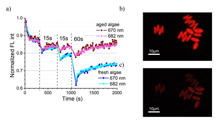
When exposed to blue light, chlorophylls fluoresce brightly in the red region of the spectrum (Fig. 1). In samples of algae from aged population the FL intensity of chlorophylls at 682 nm and 670 nm decreased relatively less than in samples of algae from fresh population (Fig. 1a). Additional irradiation with a blue diode caused short-term reversible photobleaching of microalgae FL that depended on the age of algae population. The bleaching of red-fluorescing algae cells was clearly observed after the exposure to a similar irradiation dose (Fig. 1b, c).
The exposures through the 100x oil immersion objective using three different excitation ranges ($\lambda_{ex}$=330-380 nm, $\lambda_{ex}$=380-420 nm and $\lambda_{ex}$=510-560 nm) allowed monitoring the step-wise decrease of intensity of red fluorescence leading to its total disappearance in algae cells. At first their red fluorescence turned into whitish in the area of a sample exposed at $\lambda_{ex}$=380-420 nm, and then – into the blue fluorescence. The green algae cells became transparent on the phase contrast images. Thus, a number of photoproducts with different fluorescence properties can be distinguish in algae cells by means of fluorescence microscopy during degradation of chlorophylls induced by the exposure to the blue light.