
In the past decade, stem cells, especially mesenchymal stem cells (MSCs), have been derived to track down and destroy malignant cells taking advantage of their tumor-tropic property [1]. MSCs are multipotent cells found in almost all adult tissues and have the ability to self-renew and differentiate into various cell types. MSCs are able to escape immune system and migrate to sites of lesions including tumors by using specific receptors and ligands to facilitate trafficking. Therefore, due to their tumoritropic migratory properties, MSCs can be used as delivery systems to transport nanocompounds directly to cancer cells [2].
Quantum dots (QDs) are fluorescent nanoparticles. Due to their unique optical properties and capability to form complexes and conjugates with bioactive molecules they can be used as theranostic nanoplatforms, combining both diagnostics and therapy. Low toxicity, resistance to degradation, long-term photostability and easily modified surface with drugs, photosensitizers or antibodies, enable them to be used as labels and therapeutic agents inside MSCs vehicles [3]. QDs have already been reported to be used for MSCs tracking [4], however the knowledge about QD-loaded migratory capacity of MSCs is still limited.
Before using MSCs as carriers of nanoparticles, optimal conditions for QDs loading in MSCs need to be established. In our study we, firstly, identified human skin-derived MSCs by the expression of specific surface molecules. Then we determined the optimal non-toxic concentration of QDs using cell viability test. Using confocal microscopy we detected rapid uptake of QDs and their localization in vesicles. Flow cytometric analysis showed time-dependent increase of photoluminescence intensity of QDs and the saturation point at 6 hours (Fig. 1). A more detailed analysis revealed correlation between photoluminescence intensity of QDs inside cells and the proliferation marker Ki67.
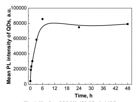
In conclusion, we optimized effective QDs concentration for labeling MSCs, found 6 hours incubation time to be sufficient to reach maximum upload of QDs and also revealed proliferation-dependent uptake dynamics.