
Gold nanoparticles (Au NPs) exhibiting unique optoelectronic properties, easy surface modification and low toxicity are one of the newest strategies in nanomedicine. Au NPs in diameter from 4 nm to several hundreds of nanometers demonstrate plasmonic absorption, therefore they can be used in phototermal cancer therapy. Due to nanoscale size comparable to the Fermi wavelength of electrons (~0.7 nm) recently synthesized photoluminescent ultrasmall gold nanoclusters (Au NCs) can easily bypass natural barriers and accumulate in cells and tissues which makes them very promising fluorescent markers. Knowledge about internalization, cellular uptake and cytotoxicity of NPs is essential before these nanomaterials can be used in nanomedicine. Unfortunately, there is still not enough information about it, therefore, more research is required. In this work cellular uptake and toxicity of 2-(N-morpholino)ethanesulfonic acid coated gold nanoparticles (Au-MES NPs) in epithelial breast cancer cells MCF-7 were investigated
NCs were synthesized according to modified synthesis protocol proposed by Bao et.al. [1] by mixing chloroauric acid solution HAuCl4 (0.29 M) and aqueous MES buffer solution (1 M, pH 6.3) to the volume ratio of 1:5. The synthesis was performed for 21.5 hours at 37 °C temperature under vigorous stirring. The synthesized colloidal solution exhibited photoluminescence at 476 nm indicating formation of ultrasmall Au-MES NCs below 1 nm in diameter [2].
To investigate uptake of photoluminescent Au-MES NCs in MCF-7 breast cancer epithelial cells laser confocal microscopy was used. The cells were incubated with Au-MES NCs (45 mg/ml) for 3, 6 and 24 hours. After 3 hours of incubation the diffuse distribution of photoluminescent Au-MES NCs within the cells was observed. After longer times of incubation photoluminescence intensity of Au-MES NCs within the cells increased. Au-MES NCs distribute evenly in total volume of a cell, including nucleus. It can be clearly seen that nanoparticles were not encapsulated in vesicles. Due to small size Au-MES NCs might have accumulated in cells via a non-endocytic pathway. Nuclear pore size for passive diffusion of molecules is about 9 nm [3], therefore particles that are smaller than the pore size, including photoluminescent Au-MES NCs. could cross envelope of nucleus.
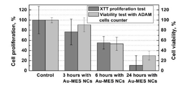
Toxicity of Au-MES NCs to MCF-7 cells was evaluated by XTT proliferation test and with ADAM cell counter. Both tests showed decreased viability and proliferation with increased time of incubation (Fig. 1). Control values were considered as 100%. ADAM viability test after 3 hours of incubation showed 91% viability. After 6 hours of incubation viability decreased to 53%. After 24 hours of incubation viability was less than 31%. XTT proliferation test after 3 hours of incubation showed 77 % proliferation. After 6h proliferation was less than 55% and after 24 hours of incubation – 19.2%. Propidium iodide dye stained dead cells were observed using confocal fluorescence microscope and confirmed results that longer time of incubation with Au-MES NCs has stronger toxic effect on MCF-7 cells. Photoluminescent Au-MES NCs show high cellular toxicity levels in breast cancer epithelial cells MCF-7.