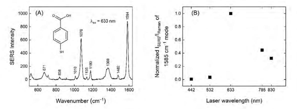
Surface-enhanced Raman spectroscopy (SERS) is an informative and non-destructive vibrational spectroscopy technique that allows for highly sensitive structural detection of low concentration analytes through the amplification of electromagnetic fields generated by the excitation of localized surface plasmons [1]. Whilst this technique has proved to be extremely useful in biomedical applications, SERS introduces some problems that need to be solved. These problems include weak reproducibility and contamination by organic materials such as reagents and stabilizers which are used for the chemical synthesis of the plasmonic part of nanoparticles. The chemical contamination can be avoided by producing nanoparticles in a cleaner way - in this case the laser ablation technique of target material in a solution that contains low concentration of salt for better nanoparticle stability. Another obstacle that appears frequently is the 'coffee-ring' effect which occurs during the sample drying as a result of uneven material distribution [2]. To avoid this kind of problems, magnetic core (Fe3O4) – plasmonic shell (Ag, Au) hybrid nanoparticles are used. The magnetic core of the nanoparticles (Fe3O4) allows effortless spatial manipulation using magnet and results in homogeneous distribution of material on a surface. These properties permit more uniform spot-to-spot SERS intensity and higher reproducibility. Multiwavelength SERS spectroscopy was carried out on the reporter molecule 4-mercaptobezoic acid (4-MBA) which was adsorbed on the smooth gold surface to investigate the enhancement capabilities of these hybrid magneto-plasmonic nanoparticles (Figure 1). In order to evaluate SERS enhancement capability of hybrid nanoparticles we calculated the ratio between SERS and Raman intensities of 4-MBA reporter molecule at the same spectroscopic setup. We found that the highest normalized SERS enhancement occurred at 633 nm, while the 785 and 830 nm excitations were moderately high.