
Prostate cancer (PCa) is the most commonly diagnosed male malignancy and the 5th leading cause of cancer-related death in men [1]. Most tumors tend to grow slowly and are low-grade with relatively lower risk of recurrence, whereas others are aggressive and significantly affect patients' life quality. PCa prognosis is primarily based on prostate-specific antigen (PSA) testing, however, it lacks accuracy in differentiating between indolent and aggressive forms of the disease [2]. Aberrant expression of histone methylation-associated genes (HMs), like histone lysine methyltransferases (KMTs) and demethylases (KDMs), has emerged as one of the major research areas in search of prognostic PCa biomarkers [3]. Being epigenetic regulators themselves, these genes could also be regulated by epigenetic mechanisms, one of which is by small regulatory RNAs, like microRNAs (miRNAs), at the post-transcriptional level. Therefore, investigation of HM gene expression together with their potential regulatory miRNAs could provide novel insights into the epigenetic mechanisms underlying PCa progression.
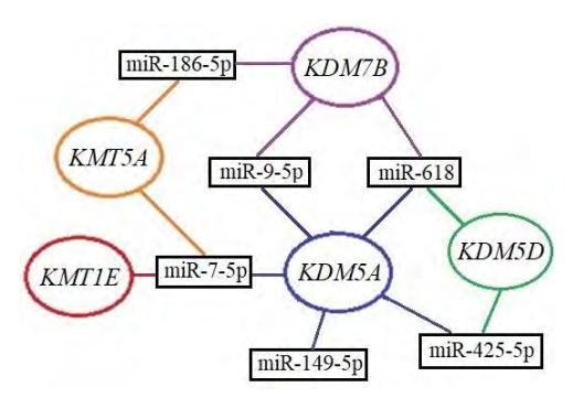
The present study was aimed at the analysis of 5 selected HM genes (KDM5D, KMT5A, KDM7B, KMT1E and KDM5A) and 6 potential regulatory miRNAs (miR-7-5p, miR-9-5p, miR-149-5p, miR-186-5p, miR-425-5p, and miR-618). In total, 186 prostate tissue samples (123 PCa, 47 noncancerous prostate tissues (NPT), and 16 benign prostate hyperplasia (BPH) samples) were included in the study. MiRNAs, which can potentially target the HM gene transcripts, were identified by in silico analysis, using data from 7 databases (Fig. 1). Expression of the selected miRNAs was evaluated in a subset of the samples (36 PCa and 6 NPT) from the same cohort. All HM genes and miRNAs were quantified by means of probe-based real-time PCR.
The results of our study revealed differences in HM gene expression among the histological sample groups. KMT5A was highly expressed in BPH as compared to both PCa and NPT (both p < 0.0001), whereas the expression of KMT1E in tumors was lower than in NPT and BPH (p < 0.0001 and p = 0.0178, respectively). Lower expression of KDM5D and KDM5A was associated with higher pathological tumor stage pT (p = 0.0342 and p = 0.0338, respectively). KDM7B had generally low expression in all samples, nevertheless, it was significantly different between pT2 and pT3 cases (p = 0.0178). No associations were found between HM genes expression and patient's age, prostate mass, and serum PSA levels (all p > 0.0500). Lower KMT5A, KMT1E and KDM5A expression was associated with biochemical disease recurrence (BCR; p = 0.0172, p = 0.0003 and p < 0.0001, respectively). Expression analysis of the selected miRNAs showed no differences between PCa and NPT and no associations were found with the HM genes expression (all p > 0.0500). Also, none of the miRNAs was associated with the BCR status (p > 0.0500).
In summary, the present study revealed significant expression differences of KMT5A, KMT1E and KDM5A in prostatic tissues, as well as some associations with the advanced disease parameters, suggesting these genes as potential PCa biomarkers. The analyzed miRNAs were not associated with the putative target genes. However, further investigation in larger independent cohorts is needed to confirm our findings.