Nanodiamonds (NDs) are of great interest as promising materials with a high sensory potential for application in various fields of science and technology. Most of the exceptional mechanical, thermal and optical properties of NDs are inherited from bulk diamond or internal point defects such as lattice vacancies. Together with vacancies, random point defects (N, Si, etc.) that form various color centers have a wide range of unique properties, such as temperature-dependent PL (SiV centers) [1], spin-dependent PL (NV color centers), no blinking or photobleaching [2,3], sensitivity of color centers to radical formation and environmental chemical reactions or changes in pH values [4]. However, several properties could be additionally obtained because of specific steps of synthesis procedure or post-processing. These properties are associated with the ND's surface and can be sensitive to the changes of the external environment.
The goal of the present study was to investigate optical properties of NDs (Ray Techniques Ltd., Israel), obtained by pulsed laser ablation and post-processed to stabilize NDs in water, as well as to reveal the dependence of their properties on the structural features of NDs and their sensitivity to the pH value of water solutions. The NDs were characterized using transmission electron microscopy, atomic force microscopy, ATR-FTIR and Raman, steady-state and time-resolved fluorescence spectroscopies.
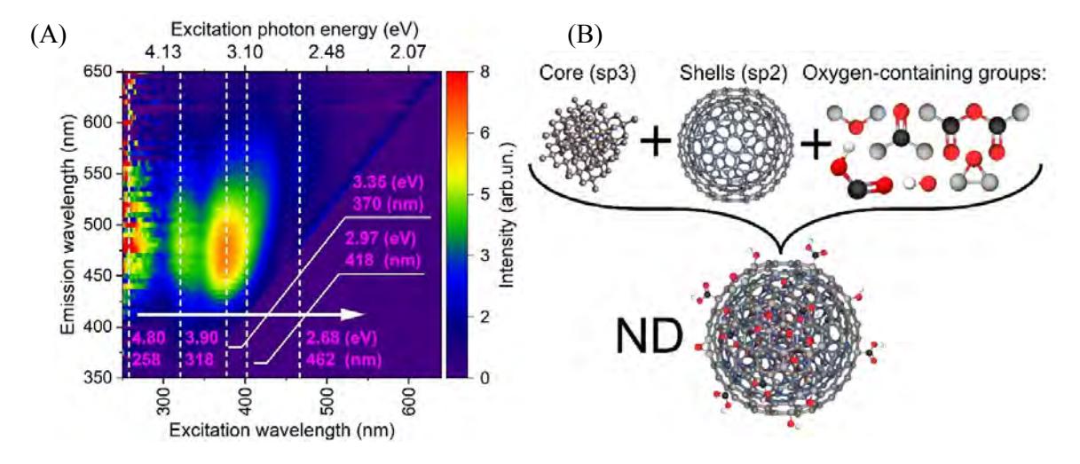
ND suspensions demonstrate a complex PL behavior which depends on the excitation wavelength, as it follows from Fig. 1 (A). The PL spectra were fitted with several Gaussian profiles allowing one to assume the presence of several PL centers in NDs. The positions of the PL maxima are slightly redshifted when the excitation wavelength increases.
The performed spectroscopic characterization of NDs reveals that NDs comprise sp3 cubic core covered with sp2-disordered clusters having size of 0.62 nm and decorated with wide range of oxygen-containing surface groups, including hydroxyl, ketone, carboxylic anhydride and carboxyl groups, without a pronounced predominance of any of them. The model is presented in Fig. 1 (B).
In the present study the revealed individual surface PL centers of NDs were shown to be pH-sensitive, and the dependence of the PL intensity on the pH was used to propose a calibration applicable for optical determination of the pH values in water solutions.