Recording of the fluorescence emitted from chlorophyll molecules is a frequently used tool to monitor photosynthetic performance, as photochemical reactions and emission are in direct competition for excitation energy. Therefore, changes in the photosynthetic rate will cause complementary changes in the intensity of the emitted fluorescence [1]. However, various factors affect emission signals in a similar way. Further investigation of fluorescence parameters is therefore needed to improve their diagnostic potential in practical applications.
Pairing an optical fiber system with a controlled stepper motor enabled registration of autofluorescence signals along Characean alga Nitellopsis obtusa internodes in 1 mm intervals. A low intensity (< 1 mW) LED light source emitting at 405 nm was used for excitation. To determine fluorescence parameters best suited to monitor photosynthetic performance under conditions of an external stress, algae were immersed overnight in a 100 μM solution of 3-(3,4-dichlorophenyl)-1,1-dimethylurea) (DCMU). This compound interrupts photosynthetic electron transport [2] and should affect the recorded emission spectra. Since it is unknown whether DCMU alters the effect of other environmental factors on fluorescence parameters, the data obtained after DCMU treatment were compared with signals registered in untreated internodes kept under different lighting conditions: darkness, natural daylight, intense (photosynthetic photon flux density of 829 μmol m-2 s-1) white LED light.
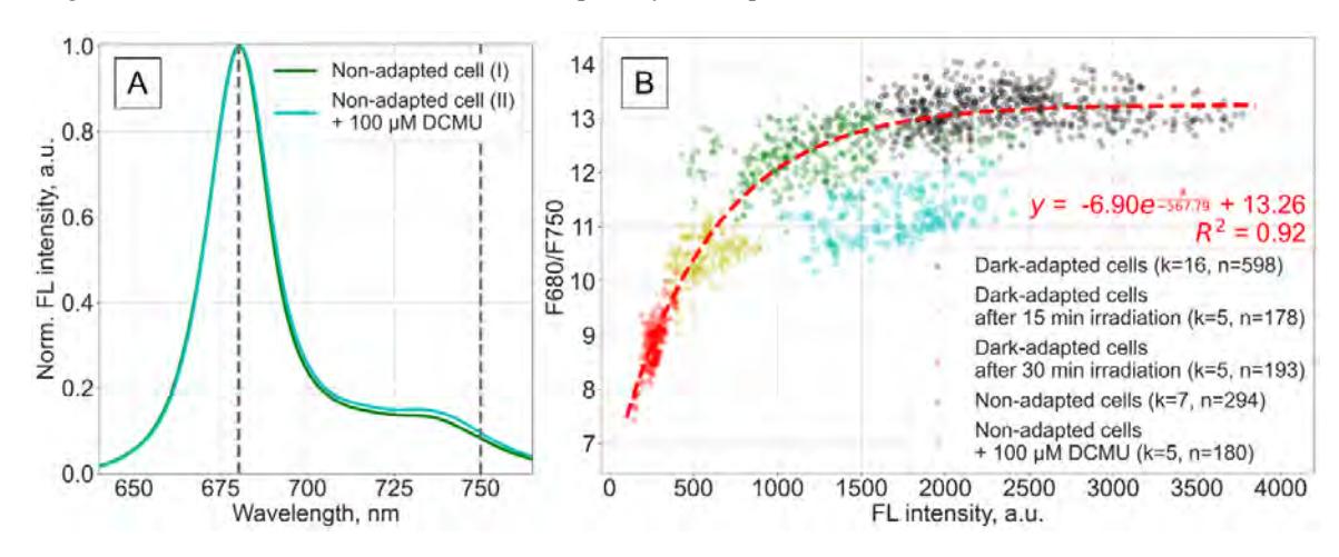
Two peaks at 680 nm and 739 nm were observed in the autofluorescence spectrum of N. obtusa (Fig. 1A). Fluorescence intensity values recorded at these wavelengths showed variations along the cell corresponding to changes in the fluorescence intensity ratio between 680 nm and 750 nm (F680/F750). These variations were observed regardless of lighting conditions. Emission spectra of algae treated with 100 μM DCMU displayed an altered shape, but the recorded intensity remained similar to that of untreated cells. Further investigation of the relationship between maximum fluorescence intensity (at 680 nm) and the F680/F750 ratio revealed a clear separation of DCMU affected algae (Fig. 1B). These results confirm that DCMU interferes with photosynthetic performance, either by affecting it in a way other than that due to the changing conditions of external lighting, or by directly altering this reaction. Therefore, the relationship found between selected fluorescence parameters provides a model for predicting light-induced changes in the emission spectra of algal cells. Nevertheless, more data are needed to confirm whether the model is suitable for assessing the effect of other environmental factors on photosynthetic performance.