
Nano-sized quantum dots (QDs) are widely used in various fields of science and engineering, including biomedicine. Although QDs may have a protective shell and surface ligands, prolonged exposure to the environment and/or light affects their structure and spectroscopic properties [1]. After decomposition, toxic metals (e.g., Cd) from the core of QDs can enter the environment, so less toxic alternatives such as Cu-based QDs are considered, despite poorer optical performance than Cd-QDs. Protein molecules can coat QDs and improve their spectroscopic properties [2], however, more research is needed to understand whether the mechanism of action of algae is similar to proteins and how they both affect the photostability of QDs.
In this work we studied the optical density (OD) and photoluminescence (PL) spectra of CdSe/ZnS-COOH (Invitrogen, USA) and CuInS/ZnS-COOH (Nanooptical Materials, USA) QDs in a model biological medium containing bovine serum albumin (BSA) or unicellular algae Scenedesmus quadicrauda when exposed to light. Samples of QDs (concentration of both QDs was 4 nM) were prepared in deionized water (dw) with BSA (concentration 100 µM) and in MWC algae growth medium with unicellular algae (concentration about 105 cells/ml). The samples were held in a 12-well plate during light irradiation, and collimated violet LED light (404 nm ± 9 nm, 30 mW/cm2) was directed at the sample from a distance of 8 cm. In all media, fractionated exposure was given at intervals of 1.8 J/cm2 and 3.6 J/cm2 to achieve a partial cumulative dose of up to 36 J/cm2.
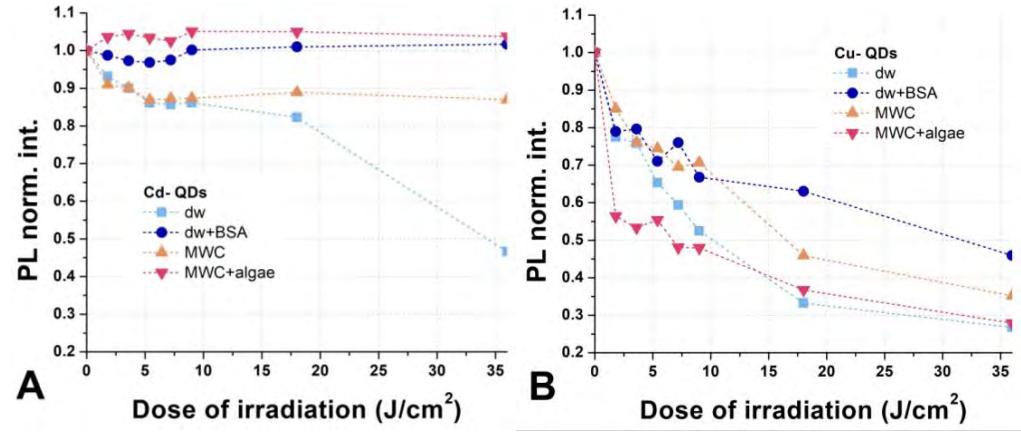
Cu-based QDs samples had a higher initial OD than Cd-based QDs, but Cd-based QDs showed a higher PL quantum yield, a narrower and more intense PL spectrum. The presence of BSA in the medium led to a higher OD and PL intensity of Cd- QDs, as well as to a bathochromic shift of PL spectrum, while in the samples with algae, there was no decrease in the PL intensity and no spectral shift. A decrease in the OD and the PL intensity and the absence of spectral shifts were registered in the samples of Cu-QDs both with BSA and with algae. After exposure to light, the spectroscopic properties of Cd-QDs were more stable in all studied media compared to the samples with Cu-QDs (Fig. 1). The presence of BSA increased the photostability of both QDs: BSA practically protects Cd-QDs from a photoinduced decrease in the PL intensity, and the decrease in the PL intensity in Cu- QDs samples was slower than in the dw sample without BSA. Although the MWC medium increased the photostability of the spectroscopic properties of both QDs compared to dw, the decrease in the PL intensity of Cu-QDs with algae after initial doses was greater than in MWC medium alone. On the contrary, the PL intensity of Cd-QDs with algae slightly increased after the initial irradiation doses and remained stable during the experiment. Cu- QDs appeared to be less photostable in general. The presence of algae in the medium further reduced their photostability, while BSA slightly stabilized the PL intensity at higher exposures. In the case of Cd- QDs, the presence of BSA or algae led to increased PL intensity of the samples.