
Background. Sodium dodecyl sulfate–polyacrylamide gel electrophoresis (SDS-PAGE) has been proved to be a high efficient method to distinguish proteins with molecular masses. There are a lot of different modifications of this method to satisfy certain purposes. In this way, the technique of substrate-containing SDS-PAG was developed to examine the proteolytic activity in samples. However, there is a necessity to optimize this method for using collagen as a substrate. [1].
The aim of the research was based on finding out appropriate conditions for conducting collagen-containing SDS-PAG by altering the main such as an incubation time, degree for sample dilution, concentrations of a separation gel and collagen solution.
Materials and methods. Tris, glycine, sodium dodecyl sulfate, acrylamide, N,N'-Methylenebisacrylamide, ammonium persulfate, N,N,N',N' – tetramethylethylenediamine were used to make gels (Sigma, USA). After electrophoretic separation gels were washed in 2.5% Triton X-100 and incubated at 37 °C for 12 hours. Samples for the experiment were blood plasma of donors, who had suffered from COVID-19, with various titers of anti- SARS-CoV-2 IgG and were received from the "BIOPHARMA-PLASMA" company (Kyiv, Ukraine). During electrophoresis two types of samples were used: 1) plasma with streptokinase (ps) which was added into a plasma: 2) plasma without streptokinase (p0). In both cases, euglobulin fractions were obtained. All samples were diluted with h the standard SDS-PAGE sample buffer. [2]. Glycerine was replaced by sucrose and dithiothreitol was removed at all to preserve enzyme activity.
Results. We had analyzed an effect of changing each of the parameters on the accuracy of this test. It was determined that the mixing ps samples with treatment buffer in the ratio 1:16 forms a larger number of evenly distributed bands. In comparison, dilution of the same ps samples by 4 and 8 times led to their concentration in one part of the plate with the formation of unclear bands. Moreover, the most accurate dilution of p0 samples was 8-fold, which produced clear and evenly distributed bands.
We explored an appropriate concentration of a separation gel by its variation: 10%, 12%, 15% and 18%. It was found that only 15% resolving gel prevented migration of samples, markers and a substrate during the experiment. In addition, we examined that using collagen solution with a concentration 2 mg/ml prevented its migration.
The time of extending the electrophoretic separation after the front line of the samples exited the gel into the electrophoresis buffer was altering between two options: 10 and 15 minutes. An accommodation of markers and samples was more correct when an elongation time was 15 minutes than 10 minutes. Also, it provided better washing after staining for undigested substrate with Coomassie Blue (Figure 1).
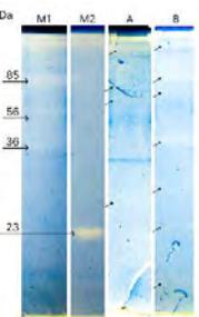
Conclusion. This study was focused on discovering set of parameters for collagen-containing enzyme-electrophoresis which will provide this test with appropriate conditions. We can state that new modification of collagen-containing enzyme-electrophoresis method which includes mixing ps samples with treatment buffer in the ratio 1:16 and p0 samples in the ratio 1:8, preparing 15% separating gel with adding 2mg/ml collagen solution and elongation of the electrophoresis for 15 minutes can be useful for different research.