
Several molecular biology protocols rely on accurate nucleic acid detection and quantification. Eventhough several developments in nucleic acid stains, labels and other detection strategies have been made [1], these rely on expensive reagents and kits. When it comes to nucleic acid staining, two important factors that must be considered include sensitivity and selectivity of the method used. We describe a sensitive and selective method for fast nucleic acid quantification based on staining with the fluorescent probe, propidium iodide (PI).
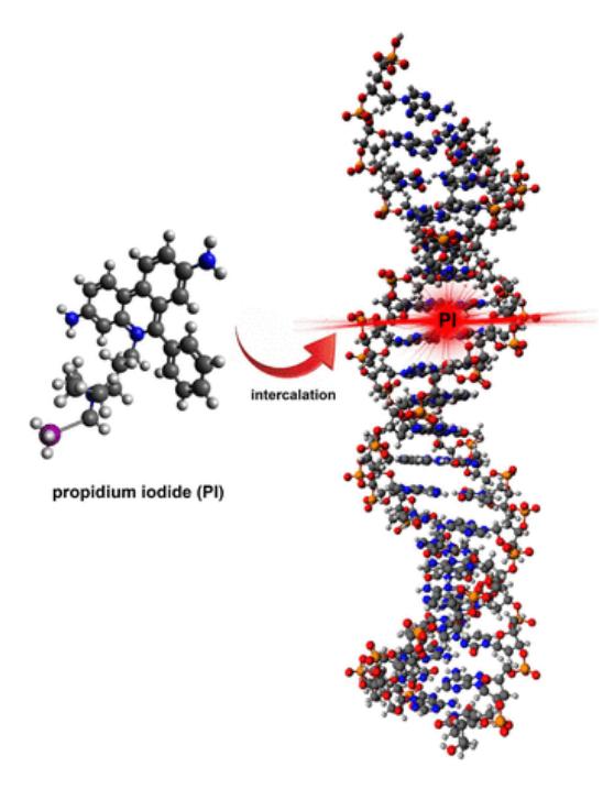
Propidium iodide is a membrane impermeant dye that intercalates between base pairs in polynucleotides. It binds stoichiometrically to nucleic acids with the emission fluorescence intensity being proportional to the amount of nucleic acid in the sample [2]. Once the dye binds to nucleic acids, it undergoes a fluorescence enhancement of approximately 20-30 fold [3]. Propidium iodide staining has been employed previously in the analysis of apoptosis using flow cytometry [2], but here we describe a method to fluorometrically quantify nucleic acids (preferentially) in solution present along with contaminating biomolecules.
CHO (Chinese Hamster Ovary) cells grown in RPMI-1640 medium supplemented with 1% L-glutamine, 10% FBS and 1% penicillin streptomycin were used as the model to study PI fluorescence emission dynamics vs PI concentration. The cells were suspended in a HEPES-based buffer and sonicated at 300-400W for 2-3s to completely disrupt the cell membrane and release their total nucleic acid (RNA + DNA) content. The results reveal a plateau in emission fluorescence intensity at PI concentrations - 40 μM/ml, 20 μM/ml, 12 μM/ml and a roughly 50% decline in emission fluorescence intensity at 1 μM/ml. We hypothesize that this decrease is a consequence of difference in selectivity of PI at low concentrations where it prefers to bind to DNA over RNA due to structural differences. Further investigations employing selective enzymatic degradation of nucleic acids will reveal the exact picture and we hope to achieve the same before the conference.