
Epithelial ovarian cancer (EOC) in most developed countries is the leading cause of death among gynaecological malignancies. The high level of heterogeneity of this cancer plays a big role in the lack of successful treatments [1]. To improve our understanding of epithelial ovarian tumour biology and behaviour it is important to adopt a suitable culture system. Usually, the majority of in vitro experiments are performed in two-dimensional (2D) cancer cell line monolayer cultures. Even though the 2D model is commonly used, it does not fully reflect the tumour complexity therefore more sophisticated models should be adopted. Cancer cell microstructures formed as three-dimensional (3D) spheroids closer represent the main characteristics of in vivo tumours, such as cell-cell interactions, hypoxia and pH rate, exposure to nutrients and metabolites, and gene expression profiles [2].
In our study, we aimed to adopt and optimize the 3D culture conditions of four different EOC cell lines: A2780, SKOV3, COV362, OV7. Next, we compared the gene and protein expression profiles between 3D and widely used 2D cell culture models by performing flow-cytometry (FACS) and analyzing the expression of genes related to stemness properties, epithelial-mesenchymal transformation and interaction with the immune system by quantitive real-time PCR (qPCR).
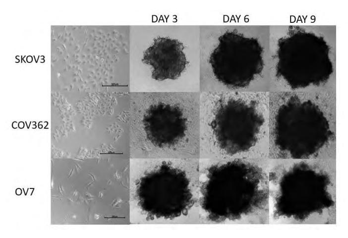
EOC spheroids were measured and observed in different growth conditions and at a different seeding density as a starting point (Fig. 1). We noticed that the A2780 cell line formed no spheroids in our culture conditions and as such this cell line was excluded from further analysis. After optimization when the best cell count and conditions were determined, protein and gene expression profile evaluation was performed. We found out that the OV7 cell line had the upregulated TGF-${\beta}$ pathway and epithelial-mesenchymal transformation-related genes. Next, we compared the gene and protein expression in respective 2D and 3D cultures, where we found substantial differences.
In conclusion, for each cell line, we characterized its gene and protein expression profile in 2D and 3D culture conditions.