
The urethra is an important component of the urinary system that facilitates the passage of urine from the bladder to the outside of the body. Effective repair of damaged urethra is essential for restoring normal urination function and preventing long-term complications[1]. Current treatment of urethral damage involves surgical intervention, with recurrent treatment often necessary. To improve treatment, we set out to use 3D printing to manufacture an artificial urethral tissue which would be suited to a patient's individual needs and would be optimal for cell proliferation, migration and differentiation into healthy urethral tissue.
Gelatin methacrylate (GelMA) and silk fibroin (SF) are two biocompatible and biodegradable polymers that have been utilized in the development of hydrogels for biomedical applications where they can serve as artificial tissues[2]. GelMA is denatured collagen modified with methacrylate, it is comercially accessible and can undergo a photoinitiated radical reaction to form a tough 3D structure[3]. Polymerized GelMA is a solid but it is not rigid enough to implant into tissues during an operation. For this reason, it was decided to combine the GelMA with silk fibroin, which when denatured forms beta-sheets and can therefore make the whole composite mechanically stronger.
The first step in creating the hydrogel was optimising the printing of the GelMA-SF construct without any cells. The workflow of printing the hydrogels consists of firsly mixing GelMA and SF, then sonicating the mixture to denature the silk fibroin, then performing the 3D printing into a non-newtonian fluid and finally using UV light to polymerize GelMA-SF into a firm, tubularized structure.
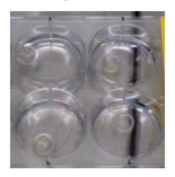
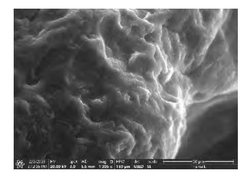
To achieve this, various component combinations of the GelMA-SF mixture and the parameters for 3D printing itself were tested. Ultimately, we found that GelMA and SF should be mixed with 10% and 5% concentrations, respectively, to consistently successfully print GelMA-SF hydrogels (fig 1.). To analyze the surface (fig 2.) of the printed construct SEM (scanning electron microscopy) was used. The acquired images show that the hydrogels' surface is uneven and has a large surface area both of which are optimal for cell migration and proliferation.
The next step of the project is to print a cell-laden GelMA-SF hydrogel. RASC (rabbit adipose stem cells) and RBMC (rabbit buccal mucosa cells) primary cells were seeded onto the hydrogel and differentiated towards the epithelial lineage for 2 weeks. Induced and spontaneus epithelial differentiation was evaluated by measuring gene expression levels of epithelial differentiation markers Ck14,Cadh1 and Ocln, and our results showed that both RASC and RBMC cells exhibit reduced expression of Ck14 after 2 weeks of differentiation on GelMA-SF hydrogels, which is a sign that the stem cells succesfully became non-proliferative epithelial cells[4].
To summarize, our results show that 3D bioprinting of cell-laden GelMA-SF hydrogels with subsequent cell differentiation into epithelial tissue could be a promising method for manufacturing artificial urethral tissue. In vivo experiments will be performed to further evaluate our findings.