
Mechanobiology is becoming well established field of science, with ever more research outlining the importance of mechanical properties of cellular environment in controlling cell fate [1], [2]. It has been shown that substrate stiffness influences cell migration, orientation and proliferation, with certain cell lines having certain preferences [3]. Here we present a simple and effective approach of creating Youngs modulus microscale patterns in a biological range, on soft biocompatible polyacrylamide hydrogel substrates. In this work we used photopolymerization of polyacrylamide enriched with methacrylic acid as the key mechanism for pattern generation. By polymerising samples in 2-step approach we were able to create zones that had higher degree of crosslinking, because of longer UV light exposure time (Fig. 1.). We were able to create this structured illumination by employing inverted microscope with “Optigrid” structured illumination module, by exchanging mask within this module with a custom one. Overall, 2 different masks were tested, which allowed for stiffness patterning of 10 µm width lines and 40 µm diameter disks. Samples had higher Young’s modulus values in exposed zones (in ranges from 10 kPa to 90 kPa for different samples) as well as some degree of elevation (up to 4 µm for line pattern and more than 10 µm for disks), confirmed via AFM measurements. Inclusion of methacrylic acid allowed for surface patterning with ECM proteins by binding primary amines to carboxy group present in hydrogel [4]. Such hydrogels have been used for cell attachment experiments, in which cells showed different morphology based on Young’s modulus value of their attachment zone. We believe that such hydrogel platforms could play a role in further mechanobiological advancements. 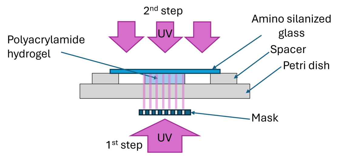
BIOCHIPS WITH MICROSCALE SURFACE STIFFNESS PATTERNS FOR MECHANOBIOLOGICAL APPLICATIONS
Andrius Pečiulis1, Tomas Rakickas1, Tadas Jelinskas1, dr. Martynas Gavutis1
1 Lithuania, Vilnius, Center for Physical Sciences and Technology, Department of Nanoengineering, LT-02300
[1] N. Gjorevski et al., “Tissue geometry drives deterministic organoid patterning,” Science (1979)., vol. 375, no. 6576, Jan. 2022, doi: 10.1126/science.aaw9021.
[2] M. T. Alameda, M. R. Osorio, J. J. Hernández, and I. Rodríguez, “Hierarchical micro-nano topographies to control Bacteria and mesenchymal stem cells biological responses,” Sci. Rep., vol. 15, no. 1, Dec. 2025, doi: 10.1038/s41598-025-08415-2.
[3] F. Milos and A. del Campo, “Polyacrylamide Hydrogels as Versatile Biomimetic Platforms to Study Cell-Materials Interactions,” Dec. 03, 2024, John Wiley and Sons Inc. doi: 10.1002/admi.202400404.
[4] M. Gavutis et al., “Biochip Surfaces Containing Recombinant Cell-Binding Domains of Fibronectin,” Coatings, vol. 12, no. 7, Jul. 2022, doi: 10.3390/coatings12070880.