
Biofilms contribute to approximately 80% of chronic or recurrent infections and exhibit high tolerance to antimicrobial therapy, often 10–1000 times greater than planktonic cells [1]. Rapid detection of biofilm growth is essential for effective treatment; however, conventional microbiological methods are time-consuming, labor-intensive, and often disruptive [2]. Electrochemical techniques offer a fast, non-invasive alternative, enabling continuous, real-time monitoring of bacterial activity at electrode surfaces [3]. The aim of this study was to evaluate biofilm formation of P. aeruginosa, S. aureus, and their mixed cultures, and to assess the influence of drugs (i.e., antibiotics) on bacterial electrogenicity.
To monitor biofilm growth, chronoamperometric measurements were performed using a three-electrode system consisting of a carbon cloth working, an Ag/AgCl (3 M KCl) reference, and a Ti foil counter electrodes, respectively. Biofilms were grown directly on the electrode surface. The chronoamperometry was used to study biofilms’ electrogenicity, which reflects the main stages of bacterial growth, including lag, log, and the stationary phase as presented in Fig. 1 A. In case with P. aeruginosa-based biofilms, the collected current increased from 0 µA to 1 µA within 24 h, indicating extracellular electron transfer between bacterial cells and the electrode surface. Mixed-species biofilms produced 2.5-fold higher current than monocultures. In the presence of antibiotics, the observed current profile had reached a plateau at 5 hours and then decreased to almost 0.0 µA, indicating their complete metabolic suspension Fig. 1 B. These findings were supported by standard microbiology assays and confocal microscopy investigations. 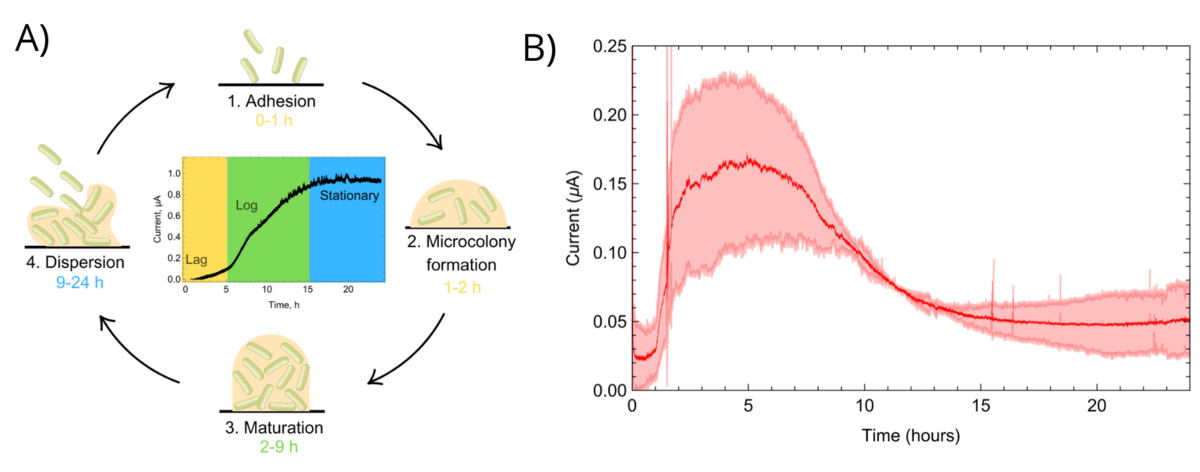
Overall, the results demonstrate that chronoamperometry enables rapid, real-time and non-invasive monitoring of biofilm formation and their susceptibility to antibiotics, providing a simple electrochemical platform for assessing biofilms’ electrogenicity and antibiotic efficacy.