
Retinoids are chemical compounds related to vitamin A (see Fig. 1) which perform various vital functions in the human body. For instance, retinol is crucial for cell growth and differentiation, while the isomerization of 11-cis-retinal to all-trans-retinal and the subsequent restoration of 11-cis-retinal during the retinoid cycle allows us to see [1]. Spectroscopic methods are frequently used for investigating the structure of such compounds, which is useful for broadening the field of applications. While experimental methods provide us with accurate results, theoretical calculations complement experimental data by offering insights about the processes being investigated on a molecular level. Various carotenoids, which are similar in structure to retinoids, were successfully studied computationally in terms of vibrational and electronic spectra using density functional theory (DFT) [2]. However, attempts to study retinoids in common solvents, especially regarding different cis-trans isomers, in a similar manner have so far been unsuccessful, despite existing experimental results [3].
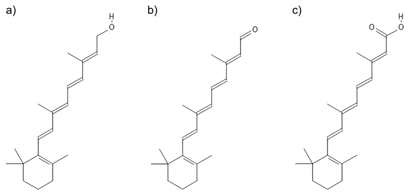
The primary goal of this study was to properly evaluate the electronic absorption energy maxima of retinol and retinoic acid in organic solvents. Molecular dynamics (MD) simulations were performed for systems containing 1 molecule of either retinol or retinoic acid dissolved in 1000 molecules of ethanol. For each compound, 2 systems were analyzed with different cis-trans isomeric forms, as the absorption maxima of retinoids are dependent on molecular configuration. Quantum mechanics/molecular mechanics (QM/MM) calculations, as well as DFT methods, were then perfromed for various configurations of each system, obtained through the MD simulations. It was found that the calculated excitation energy maxima of both retinoids depend on the description of their molecular environment – modelling the surrounding molecules with a polarizable force field yields lower values, when compared with a non-polarizable force field. Further calculations were performed, leading to the conclusion that accounting for van der Waals interactions does not have a meaningful impact on the results.